
Zebris Jaw Tracking in Digital Dentistry: Measurements, Clinical Uses and Diagnostic Limits
What can a jaw-motion recording tell us when a patient has pain, clicking or limited opening? Zebris systems can document mandibular movement and support selected digital dental workflows. A movement trace does not, by itself, establish the cause of pain, show an articular disc or validate a treatment. The useful clinical distinction is between measuring movement, identifying disease and demonstrating patient benefit. The manufacturer describes tracking as an additional assessment method; invasive decisions require other diagnostic evidence. JMA-Optic instructions, §§2.1–2.2
Which zebris system is being discussed?
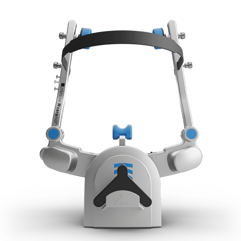
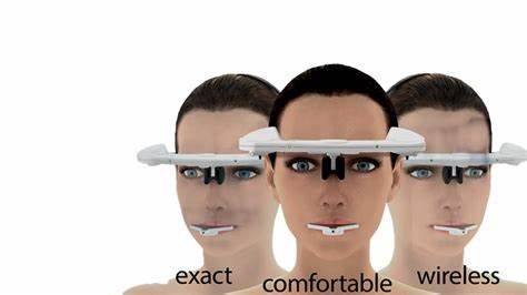
The product name matters. JMA-Optic records mandibular movement using optical triangulation. The separately documented JMAnalyser+ BT and JMT+ systems use ultrasonic transmitters and receivers. Results from an ultrasonic JMA study should therefore retain that device description rather than being presented as validation of JMA-Optic. WINJAW+ is analysis software, not a separate occlusal-force sensor. Optic hardware manual, p.8; JM systems manual, pp.12–13
The public documents checked for this article were the JMA-Optic hardware manual R3.0.2, the JM systems hardware manual dated 19 May 2026, and WINJAW+ 5.0 R1, dated 14 April 2026. The manufacturer directs users to the manuals supplied within their software for the latest applicable instructions. Check the installed hardware, software and licensed modules before transferring any workflow or specification. Official downloads
What is actually measured?
A head-mounted reference and a mandibular attachment allow the system to record the lower jaw's movement. Software then expresses selected points, paths and relationships within a defined coordinate system. A displayed condylar point is not a direct image of the entire condyle, disc or joint space. In WINJAW+'s Electronic Position Analysis, the displayed positions refer to the previously determined axis; changing the reference position or plane changes the interpretation of the report. JM systems manual, pp.7, 13; WINJAW+ manual, pp.100–102, 118
Measurement specifications are also model-specific. For example, the JMA-Optic manual lists occlusal measuring accuracy of ±0.05 mm in x, y and z within a 15 mm range of motion. The JMAnalyser+ BT/JMT+ manual lists ±0.1 mm in y and ±0.2 mm in x and z within a 15 mm range. These manufacturer specifications are not sensitivity or specificity for diagnosing TMD. Optic manual, p.18; JM systems manual, p.12
WINJAW+'s Function and Digital Occlusion module combines movement recordings with digitized dental models. It displays model contacts, contact frequency and the proximity of opposing surfaces. These geometric displays should not be reported as direct measurements of bite force or load inside the TMJ. Muscle electrical activity requires an additional recording modality; the ultrasonic JMAnalyser+ BT manual describes optional EMG accessories. A jaw trajectory alone does not measure muscle electrical activity. WINJAW+ manual, pp.69, 82; JM systems manual, pp.18–19
What do clinical studies establish?
Three different questions need separate evidence: whether a recording is repeatable, whether it identifies a particular disorder, and whether using it improves treatment outcomes. A favorable result for one question does not answer the others.
Gu and colleagues compared an ultrasonic JMA system with CADIAX 4 in 26 people aged 23–35 with normal occlusion and no TMD history. The study found no statistically significant difference in the reported displacement measures, while Bennett angles differed. Sagittal inclinations required a reference-plane correction for comparison. CADIAX 4 was not an absolute reference standard, and the authors explicitly limited the clinical implications. This was a measurement comparison, not a diagnostic-accuracy trial in patients with TMD or a trial of improved restorations. Gu et al., 2025
Zhang and colleagues retrospectively studied 74 adults, comprising 148 joints classified by MRI as having no disc displacement, displacement with reduction or displacement without reduction. Ultrasonic JMA recordings and separate surface EMG measurements differed between some groups. The study investigated group differences and correlations; it did not establish a universally applicable diagnostic threshold or test whether tracker-guided treatment improved outcomes. Small subgroups, a selected clinical population and unadjusted pain/osteoarthritis factors limit interpretation. Zhang et al., 2025
More recent evidence is promising but still specific. Xue and colleagues enrolled 40 patients with MRI-confirmed disc displacement without reduction, including 20 bilateral and 20 unilateral cases, and 20 healthy controls. In their analysis distinguishing bilateral cases from controls, sagittal opening symmetry had an AUC of 0.901 (95% CI 0.806–0.996). Participants were young: mean ages were about 18 years in the DDWoR group and 20 years in controls, and the case groups included adolescents. This was a small, single-center, cross-sectional case-control study using ultrasonic JMAnalyser+, with thresholds derived in the study sample and no reported external validation. Its result cannot be extended to all TMDs, malocclusion, other device models or treatment success. Xue et al., 2026
How should a trace fit into a TMD assessment?
Begin with the patient's history, pain and functional limitations, followed by an appropriate examination and differential diagnosis. The DC/TMD framework combines physical diagnoses with assessment of pain-related disability and psychosocial factors. Its validation supports common pain-related diagnoses; it also identifies limitations in clinical diagnosis of several intra-articular disorders. An unusual trajectory is an observation to explain in that assessment, not a disease label. Schiffman et al., 2014
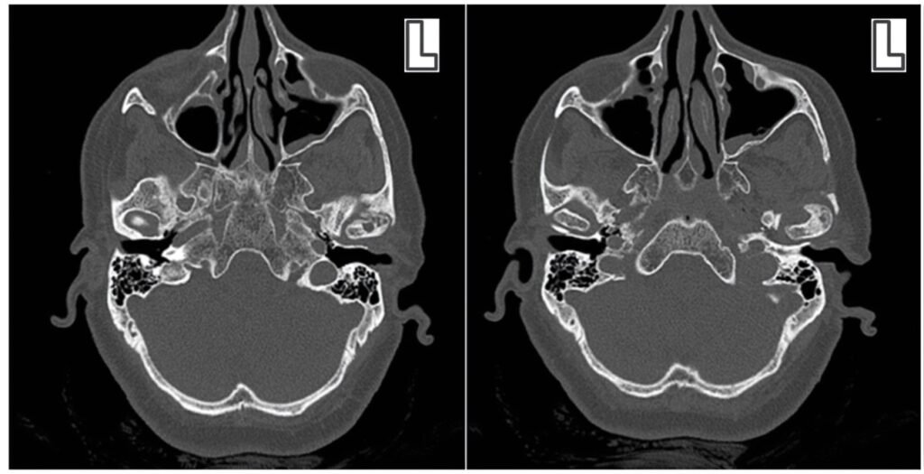
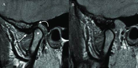
Tracking does not directly demonstrate disc displacement, degenerative bone change, reduced joint space or disc compression. When a definitive structural diagnosis would change management, the DC/TMD recommendations distinguish MRI assessment of disc displacement from CT assessment of degenerative joint disease. They also identify an exception: disc displacement without reduction with limited opening has useful clinical diagnostic validity, while alternative causes of restricted opening still need consideration. Imaging should answer a clinical question rather than automatically accompany every tracing. Schiffman et al., 2014
MRI is not inherently limited to static pictures. A feasibility study recorded real-time TMJ MRI in 40 volunteers, demonstrating disc position and the disc–condyle relationship during opening and closing. That establishes the existence of dynamic MRI techniques, not a requirement to use them routinely or evidence that either modality is universally superior. Krohn et al., 2016
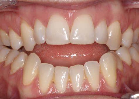
Open bite, deep bite and crossbite describe dental relationships that need clinical assessment. A movement or contact display does not establish that such a relationship caused the patient's TMD, nor does it measure joint loading. NIDCR notes that current evidence does not support a strong link between malocclusion and TMD and that irreversible bite-changing treatments have insufficient evidence as TMD treatment. Do not turn a colored contact map into a reason to grind teeth, place crowns or reposition the jaw. NIDCR: TMD causes and occlusal treatments
Can jaw tracking verify treatment success?
Repeated recordings can document changes in the measured movement task. Improvement in a trace is not, on its own, proof of pain relief, restored disc anatomy, reduced joint loading or a successful rehabilitation. Record the patient's symptoms and everyday function separately, and distinguish those outcomes from the device measurements.
A small clinical study by Pang and colleagues illustrates the distinction. Sixteen patients with TMD were assessed before and after three months of anterior repositioning appliance treatment, with 16 healthy volunteers as controls; maximum opening changed after treatment. This design does not isolate the added value of JMA-guided care against otherwise comparable care without tracking. It does not justify applying that appliance protocol to every patient with an abnormal trace. Only the abstract and publisher-displayed study excerpts were available for this review. Pang et al., 2025
For restorative work, the manufacturer supports transferring movement and articulator parameters to compatible CAD/CAM workflows. This is a documented function, not a guarantee of fewer adjustments, better longevity or resolution of headache. Clinical and laboratory teams should keep the registration method, reference plane and intended use with the transferred record, and verify the restoration clinically. Optic manual, §2.1; Gu et al., 2025
How were jaw-motion records used in a real rehabilitation?
A published case shows how jaw-motion records were used to transfer and cross-check a planned jaw relationship during rehabilitation for severe tooth wear. The seven steps below connect the clinical problem with digital design, manufacture and delivery. The measurements were interpreted alongside clinical examination and patient adaptation. Lou et al., 2026.
The source names zebris Medical GmbH as the jaw-motion analyzer manufacturer. Its exact JMA model is unspecified, so this case cannot be assigned to optical JMA-Optic or ultrasonic JMAnalyser+. It cannot establish a standalone TMD diagnosis, an ideal jaw position or the added effectiveness of a particular tracking system.
This is the same published patient case discussed from the muscle-recording perspective in the DDS surface EMG article. It is not a second patient or a DDS-treated case. Here, the teaching focus is the transfer and clinical checking of jaw-motion records.
Use the numbered or previous/next links, or swipe sideways. Select the vertical-list option to read down the page. All steps and captions are available without JavaScript. Larger-view links show the available reviewed image size; small printed values are not used for numerical interpretation.
Step 1 of 7 · Clinical assessment
Establish the restorative problem
First stepNext: step 2
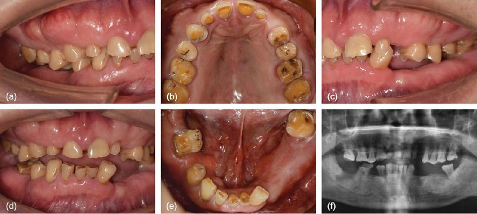
The report describes a 61-year-old man with extensive tooth wear, missing teeth and reduced occlusal vertical dimension. He was clinically asymptomatic at the temporomandibular joints despite mild imaging changes. The restorative indication arose from the clinical and radiographic assessment.

Figure 1a–fPretreatment intraoral views and the panoramic radiograph document the restorative problem before jaw-motion data are interpreted. The panoramic subpanel is included for case context only; this reduced overview is not for diagnostic radiographic reading or measurement. Source and licence. DDS teaching point. For the dentist, keep the restorative problem, symptoms and imaging findings separate from the movement measurement.
Evidence limit. This is a restorative rehabilitation case, not evidence that jaw tracking diagnosed or cured TMD.
Step 2 of 7 · Clinical and digital records
Transfer movement records into planning
The authors used centric-relation records and Gothic arch tracing, then a mechanical facebow and physical articulator for cross-checking. They report that the JMA recorded asymmetric trajectories during opening, protrusion and lateral movements. Movement data were exported as XML and imported into a virtual articulator.
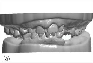
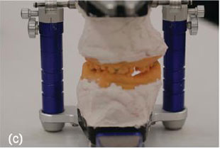
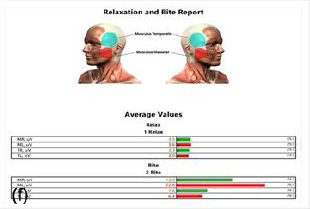
The selected panels show dental models, physical articulation and a separate averaged EMG report. JMA acquisition and XML export are source-reported steps, not pictured here. The paper names zebris Medical GmbH as the JMA manufacturer but gives no exact model.

Figure 2aInitial STL dental models provide a digital representation of the dentition. This panel does not display the jaw-motion recording. Source and licence. Figure 2cMounted casts provide the physical-articulator reference used alongside the digital records. Source and licence. 
Figure 2fThe separate surface EMG display shows rest/bite average-value bars. It is not a jaw-motion trace or a before-and-after comparison. Source and licence. DDS teaching point. For the laboratory, identify which record represents movement, which represents the jaw relationship and which comes from a separate measurement modality.
Evidence limit. Do not assign this case to JMA-Optic or JMAnalyser+. Figure 2f contains averaged EMG bars, not a jaw-motion trace, raw EMG waveform, serial comparison or numerical treatment threshold.
Sections 2.2, 2.3.1 and 2.3.2; selected Figure 2a/c/f; PDF pages 3–4
Step 3 of 7 · Digital design, manufacture and clinical insertion
Test the planned relationship clinically
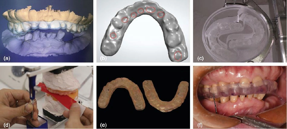
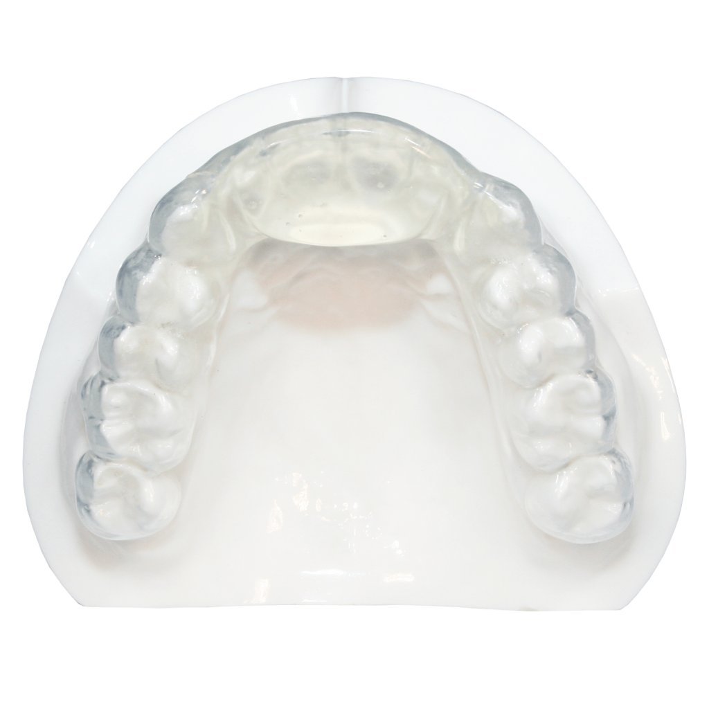
A digitally designed, milled occlusal stabilization splint was checked on the physical articulator and inserted clinically. The authors report acceptable comfort and clinical mandibular stability after three months. Progression depended on the clinical assessment; serial EMG was not systematically repeated at this stage.
The wider rehabilitation included implant treatment. The source reports second-stage surgery after six months of osseointegration; those surgical stages are not pictured here.

Figure 3a–fSplint CAD and virtual contacts are followed by CNC milling, articulator verification, the finished splint and clinical insertion. The overview identifies workflow stages; small contact markings are not a basis for selecting adjustment targets. Source and licence. DDS teaching point. A transfer that looks consistent in software still requires a clinical assessment of the appliance and the patient’s response.
Evidence limit. These photographs do not establish an ideal jaw position, disc correction or muscle normalization.
Step 4 of 7 · Clinical records and physical verification
Recheck the relationship during provisionals
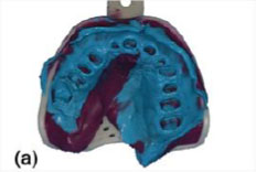
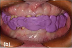
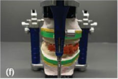
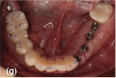
The report describes JMA re-evaluation of the therapeutic relationship. During the second provisional phase, impressions and centric-relation records were followed by cast digitization, virtual mounting, CAD and physical-articulator verification. The selected panels show the clinical records and physical check; the omitted digital panels are not reconstructed.
The source reports assessment of fit, esthetics, phonetics, comfort, temporomandibular and muscle symptoms, and restoration stability. Occlusal assessment was a separate measurement. The provisional adaptation period lasted approximately three months.

Figure 5aAn impression records the prepared dentition during the second provisional phase. Source and licence. Figure 5bThe centric-relation record is one of the clinical records used to transfer the planned relationship. Source and licence. Figure 5fPhysical-articulator verification remains part of the case’s hybrid workflow. Source and licence. Figure 5gIntraoral clinical verification shows prepared teeth and implant abutments; this is not an unequivocal view of a complete seated provisional. Source and licence. DDS teaching point. For the dentist and laboratory, preserve the distinction between the recorded relationship, virtual simulation and what was actually checked in the mouth.
Evidence limit. The first-provisional stage is not assigned a precise place here because the paper’s summary diagram and body differ in their ordering. Figure 5g is not an unequivocal image of a complete seated provisional.
Step 5 of 7 · Digital design and manufacture
Carry the checked relationship into definitive design
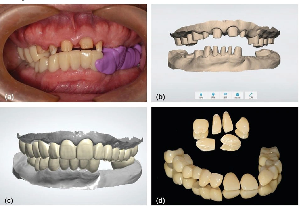
After provisional adaptation, intraoral scan data were registered to the verified interarch relationship. The source documents digital master models, full-contour CAD and manufactured zirconia restorations. No prospectively defined numerical JMA threshold determined clinical acceptability.

Figure 6a–dSectional provisional removal preserves the recorded relationship for digital master models, definitive CAD and manufactured zirconia restorations. Small interface labels are not used to infer CAD settings, dimensions or margin fit. Source and licence. DDS teaching point. For the laboratory, retain the verified relationship during model registration and design; the illustrated case does not supply a universal occlusal prescription.
Evidence limit. These images show case-specific CAD and manufacture, not proof that a tracking system selected an optimal occlusion or improved longevity.
Step 6 of 7 · Clinical delivery
Verify delivery in the mouth
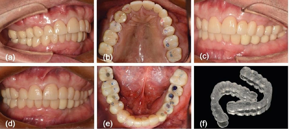
At try-in and delivery, the authors assessed occlusion with a separate digital occlusal-analysis system and articulating paper, then adjusted contacts. They delivered the definitive restorations and a protective nocturnal splint.

Figure 7a–fDefinitive restorations and the protective nocturnal splint are shown at delivery. These are not dated five-year photographs. This delivery overview does not independently verify individual contacts, restoration fit or tissue health. Source and licence. Open Figure 7a-f at a larger reviewed sizeLou et al., Figure 7a-f
DDS teaching point. Judge clinical contacts and the patient’s response independently of whether the digital transfer appeared successful.
Evidence limit. The delivery images do not document the five-year review or demonstrate the added benefit of jaw tracking.
Step 7 of 7 · Immediate digital occlusal assessment
Separate the immediate check from long-term outcome
Previous: step 6Final step
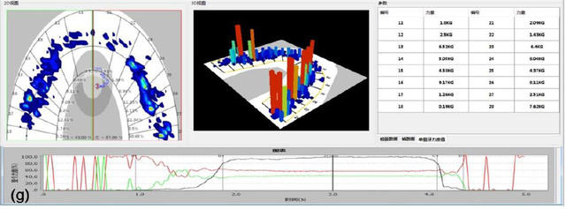
Figure 7g belongs to the delivery-stage occlusal assessment. The longer-term clinical observations are reported separately below. Neither this display nor the earlier EMG panel is a longitudinal jaw-motion comparison.

Figure 7gA separate digital occlusal assessment documents the delivery-stage contact check. It is neither a jaw-motion recording nor an EMG result. Small table and plot values are not for numerical interpretation; no threshold or measurement is read from this overview. Source and licence. Open Figure 7g at a larger reviewed sizeLou et al., Figure 7g
DDS teaching point. Report a movement finding, a contact assessment and patient benefit as separate observations; do not treat one as proof of the others.
Evidence limit. No dated five-year clinical photograph, serial JMA trace comparison or systematic serial EMG dataset is supplied for this sequence.
What was reported over five years?
Separately from the delivery images, the authors report maintained bilateral centric contacts and prosthesis integrity during five years of maintenance, including three minor occlusal adjustments. They report no implant loss, clinically diagnosed peri-implant mucositis, screw loosening, ceramic chipping, restorative fracture or visible mechanical fatigue. These are observations in one patient, not a complication rate.
Patient satisfaction was recorded with a study-specific, unvalidated scale. No dated five-year clinical photograph or systematic serial EMG comparison is supplied. The reported course cannot establish that jaw tracking caused the outcome, was superior to conventional care or validates any diagnostic threshold. Section 2.3.5 and Discussion.
What should the dentist and laboratory take from this case?
The source illustrates movement-data transfer within a staged clinical workflow. The DDS educational application is to preserve that chain of information: establish the indication, identify the recording and reference method, transfer the planned relationship, and assess the patient and restoration clinically at each relevant stage. A software display remains one part of that assessment.
Published case source and image credit. Lou C, Atashbahar M, Xu C, Liu Q. 4D Digital Kinematic Workflow for the Full-mouth Rehabilitation of Severe Tooth Wear: A 5-Year Case Report. The Open Dentistry Journal (2026), 20:e187421064592. Figures 1a–f, 2a/c/f, 3a–f, 5a/b/f/g, 6a–d and 7a–f/g. © 2026 the authors; published by Bentham Open. Reused under CC BY 4.0.
Figures 1, 3 and 6 retain the complete figure composition from the publisher PDF. Other images retain selected source panels and their printed labels; Figure 7a–f remains grouped as published. Figures 1, 3, 6 and 7a–f use resized WebP display variants with Q95 lossy encoding, plus an available source-width Q95 view. Figure 7g uses lossless WebP encoding after resizing, with a largest reviewed view of 640 pixels. The seven small Figure 2 and Figure 5 panel files retain their existing lossless WebP bytes. Selected panels are arranged in HTML, and DDS captions and teaching points have been added. No upscaling, recoloring, added diagnostic markings or diagnostic retouching was performed.
The authors report written consent for publication of the clinical details and clinical/radiographic images; the signed form was not available for this review. No patient, author or manufacturer endorsement of DDS is implied. Publisher PDF, licence on page 1 and consent statement on page 9.
Practical interpretation for the dentist and laboratory
- Define the question first: movement documentation, articulator programming and diagnosis are different tasks.
- Identify the hardware, software, reference plane and attachment method before interpreting or transferring a record. Poor fixation or recording artifacts require checking the acquisition, not inventing a pathology.
- Describe what was observed: for example, a limited or asymmetric trajectory during a stated task. Keep the structural diagnosis and patient-reported outcome as separate findings.
- Apply research to the population, device and endpoint actually studied. A group difference or a sample-derived ROC result does not establish a treatment indication.
- Use tracking alongside the clinical assessment. The manufacturer's warning remains central: invasive or patient-risking measures must not be based on tracking results alone.
These are educational interpretation checkpoints drawn from the cited manuals and studies. The value of jaw tracking lies in making movement records interpretable and transferable while keeping their clinical limits visible.
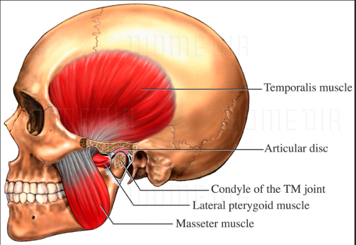
Device and anatomy illustrations
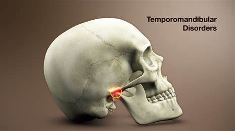
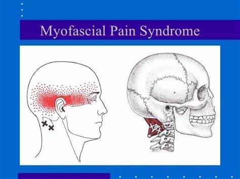
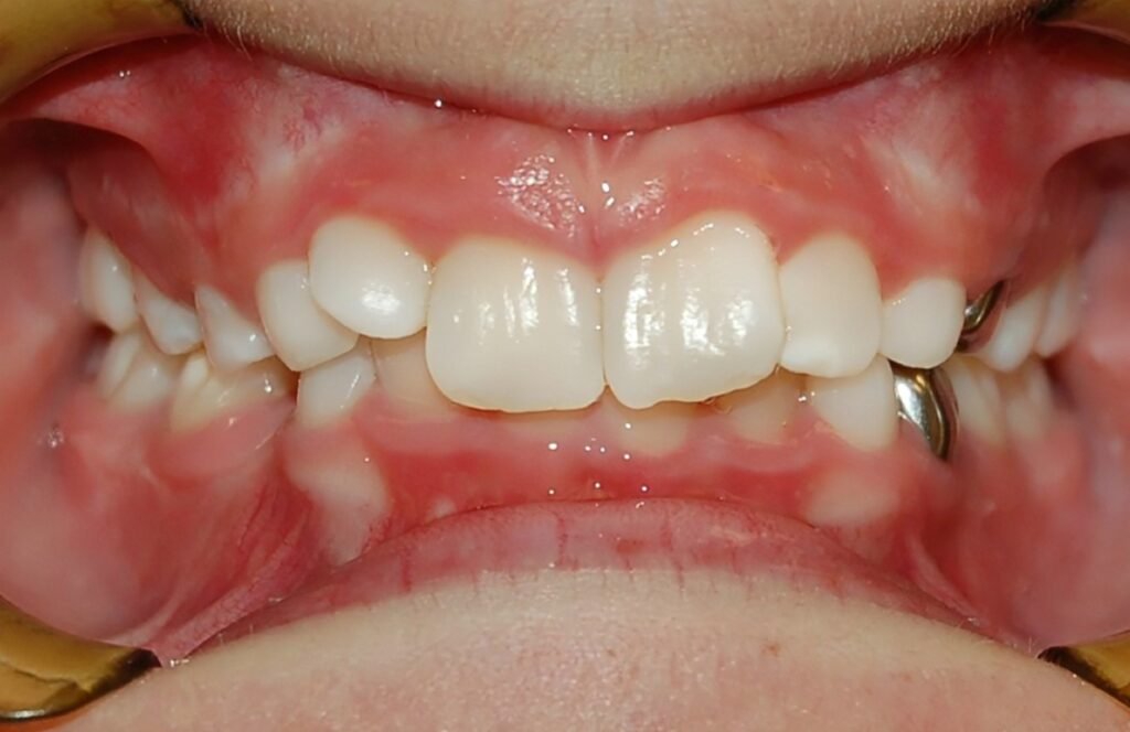
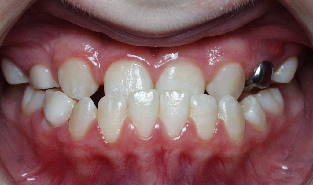
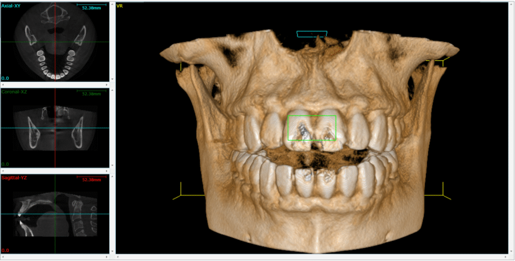
These illustrations support the technical discussion. They are separate from the sourced patient case.

WHERE DIGITAL MEETS CLINICAL
DIGITAL DENTISTRY SCHOOLOGY

Perio-Implantologist
International Digital Dentistry Speaker
Digital Occlusion Consultant
Founder of DDS
Founder of Occlusa AI Platform
 h.sharshar@ddschoology.com
Written by Dr Haitham Sharshar
Dr. Haitham Sharshar is an international digital dentistry speaker, educator, and digital occlusion consultant based in Cairo, Egypt. His work focuses on functional digital dentistry, CAD/CAM, implantology, jaw-motion analysis, and the integration of digital diagnostics into clinical treatment planning.
As Founder and Scientific Coordinator of Digital Dentistry Schoology (DDS), he has trained more than 3,270 dentists and dental technicians through university programs, international conferences, and hands-on courses. His teaching connects patient-specific diagnostic records with digital design and clinical workflows, helping clinicians and technicians understand how function and occlusion inform restorative planning.
Dr. Sharshar is a certified trainer for zebris JMA-Optic+ jaw-motion analysis and MyoWise dental EMG. His educational and consulting work brings together patient diagnostics, jaw-motion records, muscle-activity data, digital occlusion, and AI-supported workflows.
He is Founder and Clinical Director of Occlusa, an AI-supported platform for organizing clinical information and supporting clinician-led review and treatment planning. He also owns HS Dental Clinic in Cairo, where his clinical focus includes full-mouth digital rehabilitation and smile design.
His speaking and training topics include functional digital dentistry, digital occlusion, jaw-motion analysis, CAD/CAM workflows, digital implantology, and the practical integration of AI-supported tools into dental education and clinical practice.
Happy to collaborate on spreading digital dentistry.