
Dental Surface Electromyography (EMG)
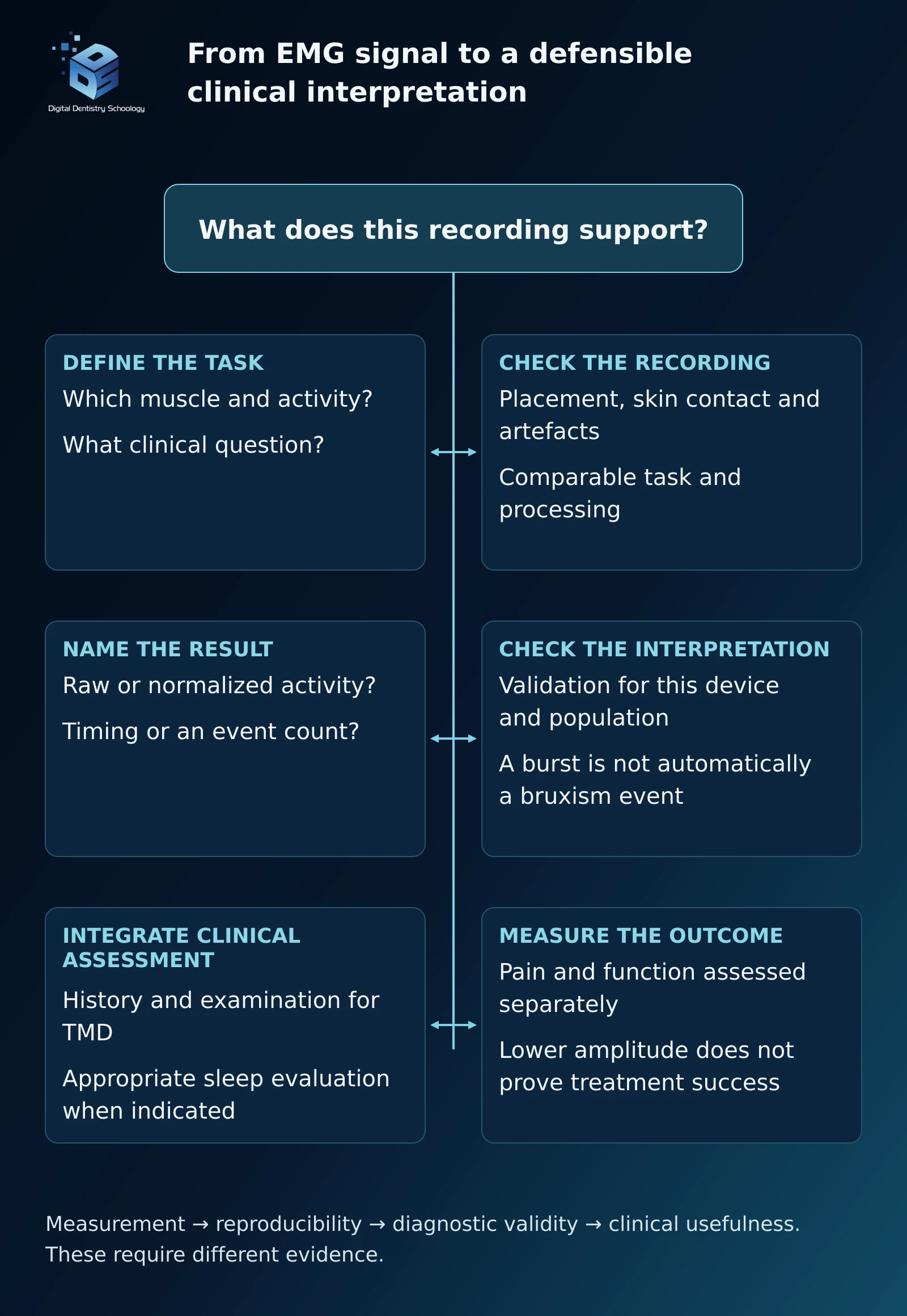
What can a dental surface EMG trace tell you, and what does it leave unresolved? An electromyography report can look persuasive: left and right muscle traces, colored indices and a change after treatment. The important question is what each result has actually measured. Surface electromyography (sEMG) records muscle electrical activity. Interpreting that activity as a diagnosis, the cause of symptoms or proof of treatment success requires additional evidence.
This DDS review follows the recording from acquisition to clinical interpretation. Its focus is dental surface EMG, particularly the masseter and anterior temporalis, rather than diagnostic needle EMG used in neurological assessment. The aim is to make the information useful without asking a muscle trace to answer questions it cannot resolve.
What does surface EMG measure?
Electrodes over superficial muscles detect electrical activity during defined conditions, such as rest, clenching or chewing. The amplitude and timing describe the signal recorded under those conditions. They do not directly measure bite force, pain or the position of the TMJ disc. Estimating muscle force from EMG requires additional biomechanical information and assumptions; the CEDE consensus describes only limited circumstances in which such estimates are appropriate. CEDE force-estimation consensus, 2024
Keep four questions separate:
- Measurement: What activity and task were recorded?
- Reproducibility: Would the same protocol give a sufficiently consistent result?
- Diagnostic validity: Does the proposed interpretation identify the condition in the intended population?
- Clinical usefulness: Does acting on that information improve an outcome that matters to the patient?
A repeatable measurement addresses the second question. It does not automatically answer the third or fourth. This distinction is the practical thread linking the measurement and clinical studies below.
Can an EMG trace diagnose TMD or an occlusal problem?
In a case-control study of 36 patients with myofascial pain and 36 asymptomatic controls, resting EMG and symmetry measurements did not reliably distinguish the groups. Controls generated higher activity during clenching. The findings do not support diagnosing an individual patient from a high resting value or a left–right difference. They also challenge the assumption that painful muscles must always produce a larger signal. Manfredini and colleagues, 2011
TMD assessment remains grounded in the history and an appropriate clinical examination. The DC/TMD framework separates muscle pain, joint pain and intra-articular diagnoses rather than treating them as one electrical pattern. Surface recordings cannot directly show disc displacement, and muscle and joint disorders can coexist. DC/TMD diagnostic framework
The same restraint applies to occlusion. An asymmetric trace does not establish that a bite discrepancy caused pain or that selective grinding will help. NIDCR reports that evidence does not support the belief that a bad bite or orthodontics causes TMD, and cautions that treatments permanently changing teeth or the bite may worsen problems. Adjustment for a separately established restorative indication is a different decision; an unvalidated EMG cutoff should not supply the indication. NIDCR
How do you make a recording interpretable?
Consistency starts before the electrodes are attached. Define the question and use the selected device’s instructions, rather than borrowing distances, filters or task settings from an unrelated system. A reproducibility study in 15 healthy volunteers used anatomical landmarks and repeated sessions to examine its masticatory-muscle protocol. That is useful measurement evidence, but it does not establish a universal normal microvolt range. Sabaneeff and colleagues, 2017
A practical recording record should identify:
- Equipment: device, software, electrode type and sampled muscles.
- Preparation: skin preparation, electrode location and spacing, and reference-electrode arrangement.
- Task: posture, rest or contraction instructions, duration and relevant effort conditions.
- Signal handling: acquisition settings, filtering or other processing, and checks for movement or contact artefacts.
- Comparison method: raw or normalized amplitude, the reference contraction used, and which conditions were kept consistent between visits.
This is an educational documentation checklist, not a replacement for a device’s operating instructions. If the signal changes after correcting electrode contact or placement, resolve the recording problem before attributing the change to disease.
Normalization expresses activity relative to a specified reference. Its suitability depends on the clinical or research question. A 2025 study of healthy young adults found electrode position affected raw potentials more than its standardized indices. That supports a measurement-consistency discussion, not a claim that a normalized score reveals an ideal occlusion. CEDE normalization consensus, Rosati and colleagues, 2025
What can EMG contribute to bruxism assessment?
Bruxism is broader than audible tooth grinding. Awake activity can include sustained or repetitive tooth contact, jaw bracing or thrusting. The 2025 international consensus describes subject-based, clinically based and device-based assessment, rather than treating these as a simple ladder of certainty. The methods provide different information about activity and its possible consequences. Verhoeff and colleagues, 2025
Portable EMG can sample jaw-muscle activity outside the clinic, but not every burst represents a bruxism event. Miettinen and colleagues compared seven recording setups in 19 volunteers. EMG-only setups could miss or overcount events compared with audiovisual polysomnography. Their study involved one scorer and specific setups, so its results should not be generalized to every current wearable. Miettinen and colleagues
What does a real misclassified recording look like?
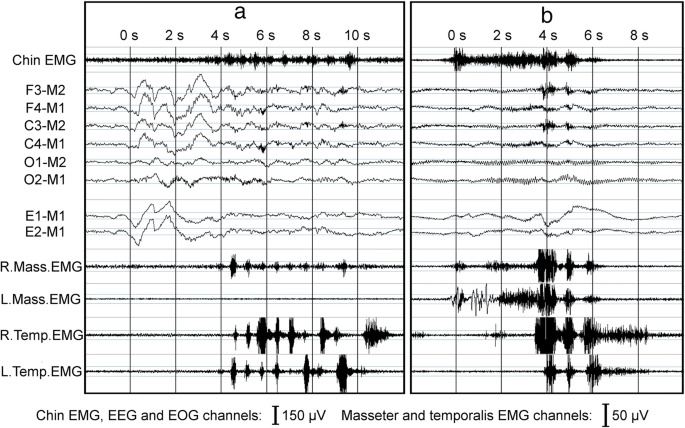
The lesson is concrete: a visible muscle burst and its event label are separate pieces of information. Ask which channels and validation support the label before accepting an automated count.
Sleep-disordered breathing needs its own assessment. In a 22-person study of a portable EMG–ECG device, interpretation was less dependable in moderate or severe obstructive sleep apnea. A muscle trace alone cannot exclude apnea or establish why an event occurred. When breathing-related sleep symptoms are suspected, an appropriate sleep evaluation is more informative than relabeling the EMG result. Portable-device comparative study, 2023
Does a smaller signal after treatment mean the patient improved?
It shows a change in recorded activity under the measurement conditions. Pain relief, comfortable function, tooth protection and long-term restorative survival are separate outcomes. They must be assessed separately rather than inferred from the graph.
Botulinum toxin illustrates the distinction. A placebo-controlled sleep-bruxism trial randomized 30 participants and analyzed 23 completers. Masseter burst amplitude fell over 12 weeks, but the occurrence of rhythmic masticatory muscle activity did not decrease correspondingly. A lower amplitude therefore did not demonstrate that the sleep activity had stopped. Shim and colleagues, 2020
In a different trial involving 100 women with persistent myofascial pain, botulinum toxin improved pain relative to saline but did not outperform an oral appliance at the final assessment. Dose-related adverse effects included temporarily reduced masticatory performance, reduced muscle thickness, and reduced bone volume in the mandibular coronoid and condylar processes. These findings do not provide a universal dental injection protocol or justify selecting and repeating injections from an EMG score alone. De la Torre Canales and colleagues, 2020
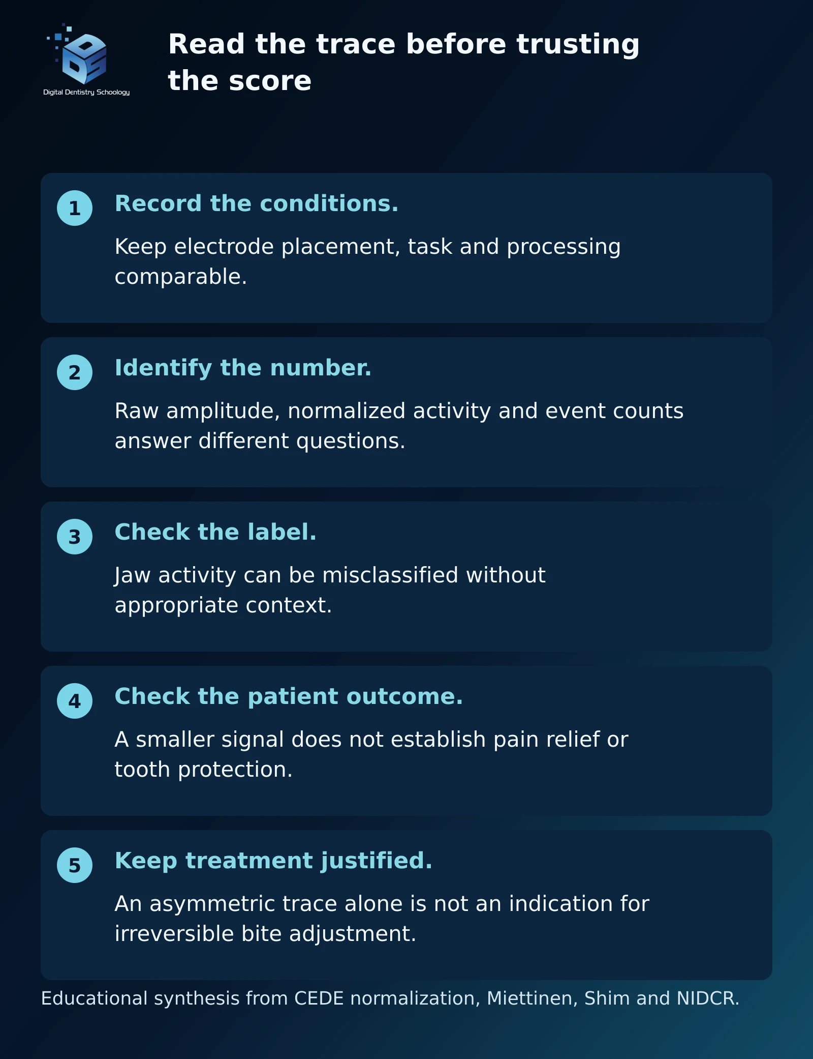
Interpretation exercise: A follow-up report shows lower masseter amplitude. Before calling it a treatment success, check whether electrode placement, task, effort, processing and normalization were comparable. Then compare independently recorded pain and function. This is a hypothetical reasoning exercise, not a DDS patient outcome. The measurement checks follow the normalization consensus; the distinction between amplitude and event occurrence is demonstrated by the Shim trial.
Where does EMG fit in an actual rehabilitation?
Where did EMG fit into an actual rehabilitation? This published adult tooth-wear case combines clinical examination, jaw-motion records, digital design and conventional checks. EMG supplied limited adjunct information early in the workflow. Progression depended on clinical findings and patient adaptation, not an EMG cutoff. Lou and colleagues’ 2026 case report is the source of all seven steps; this is not a DDS patient case.
The patient had severe tooth wear and missing teeth and was clinically asymptomatic at the TMJs. The case illustrates an adjunct recording within a wider restorative workflow. It cannot establish TMD diagnostic validity, comparative effectiveness or an EMG-led cure. The source names zebris Medical GmbH as the jaw-motion analyzer manufacturer. No exact JMA model or EMG system is established in the source. The captions therefore remain device-neutral.
For the joint assessment pathway, see the DDS guide to TMJ disc displacement. A surface EMG report cannot show disc position.
Use the numbered or previous/next links, or swipe sideways. Check “Read all steps in one vertical list” to read down the page; uncheck to return to slides. All steps and captions are present without JavaScript. Open a figure for its available native resolution; small source labels and values are not a basis for numerical teaching.
Step 1 of 7 · Clinical assessment
Start with the clinical indication
First stepNext: step 2

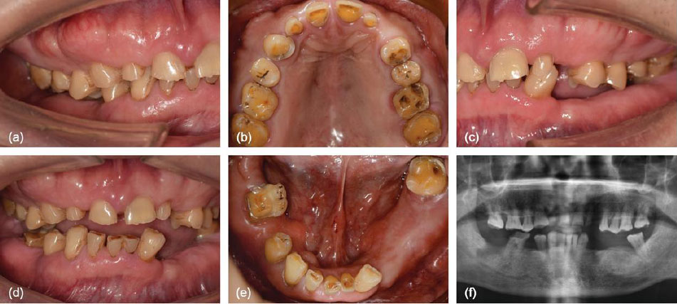
Figure 1Pretreatment intraoral views and panoramic radiograph in the published tooth-wear case. Decision and rationale. Assess the worn dentition and missing-tooth problem before selecting the rehabilitation. Clinical and radiographic findings established the restorative indication; EMG did not supply the diagnosis.
Checkpoint. Keep the documented indication separate from the muscle recording.
Evidence limit. The patient was clinically asymptomatic at the TMJs. This is not a TMD-cure case.
Step 2 of 7 · Clinical and digital records · adjunct EMG
Add EMG to the wider record
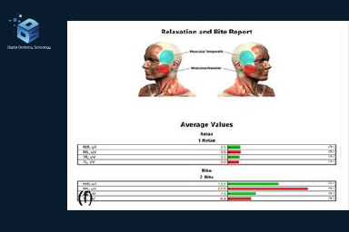
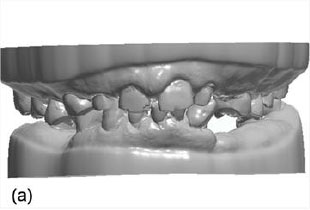
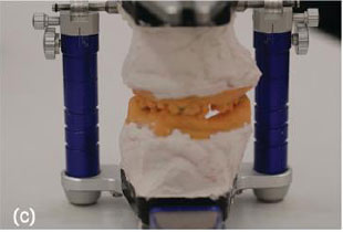
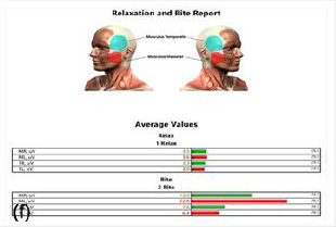
Digital dental models, physical articulation and the adjunct rest/bite average-value EMG report.

Figure 2aInitial digital dental models (STL) Figure 2cMounted physical articulator 
Figure 2fRest/bite average-value EMG report, titled Relaxation and Bite Report; not a raw serial trace Decision and rationale. Interpret the adjunct EMG record alongside jaw-relation records, jaw motion and analog verification. The authors used EMG descriptively during initial assessment; it did not independently determine treatment progression.
Checkpoint. Identify the recording type and documented task before interpreting its meaning.
Evidence limit. This is not a raw waveform or baseline/follow-up trace pair. The values are too small for reliable numerical teaching. No prespecified EMG acceptance threshold or EMG model is reported.
Source: Selected Figure 2a, 2c, 2f; sections 2.2 and 2.3.1-2.3.2; PDF pages 3-4
Step 3 of 7 · Digital design · manufacture · clinical insertion
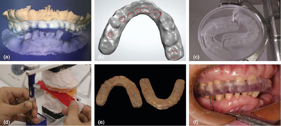
Design and test a reversible splint

Figure 3The splint was designed digitally, milled, checked on an articulator and inserted clinically. Decision and rationale. Cross-check the proposed relationship through splint design, fabrication and clinical adaptation. The report describes clinical comfort and mandibular stability as progression criteria rather than an EMG cutoff.
Checkpoint. Verify physical contacts and the patient’s functional response.
Evidence limit. Serial EMG was not systematically repeated; the photographs do not demonstrate EMG normalization.
Step 4 of 7 · Clinical provisional verification
Verify the provisional phase
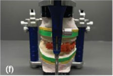
Impression and jaw-relation records, physical-articulator verification and an intraoral check during the second provisional phase.

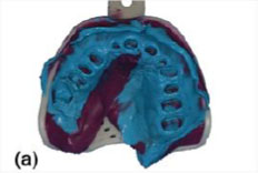
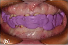
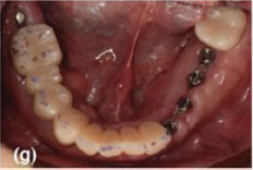
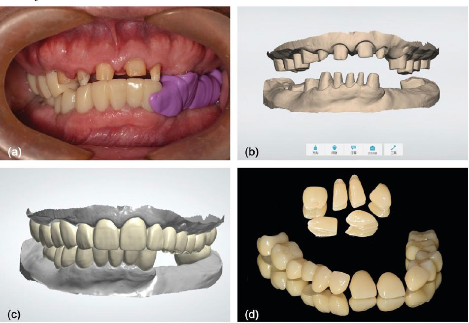
Figure 5aConventional impression after tooth preparation Figure 5bCentric-relation record Figure 5fPhysical-articulator verification Figure 5gIntraoral clinical verification view showing prepared teeth/abutments; not an unequivocal complete seated provisional Decision and rationale. Recheck records and physical/clinical consistency before the definitive restorations. The report assessed fit, comfort, esthetics, phonetics, symptoms and restoration stability during the provisional phase.
Checkpoint. Clinical adaptation and verification must accompany the digital plan.
Evidence limit. Panel 5g does not clearly show a complete seated provisional. The exact timing of the first provisional stage differs between the source body and Figure 8; no exact first-provisional timeline is asserted.
Source: Selected Figure 5a, 5b, 5f, 5g; section 2.3.4; PDF pages 4-5
Step 5 of 7 · Digital design · manufacture
Transfer the verified relationship

Figure 6The verified relationship was transferred to digital master models, definitive CAD and manufactured zirconia restorations. Decision and rationale. Preserve the verified interarch relationship while designing and making the definitive restorations. The source describes IOS/CAD transfer following clinical adaptation to the provisional phase.
Checkpoint. Cross-check the transferred relationship and restoration fit.
Evidence limit. This is a case-specific restorative workflow. It does not show that EMG prescribed the occlusal design.
Step 6 of 7 · Clinical delivery
Deliver and protect the restorations

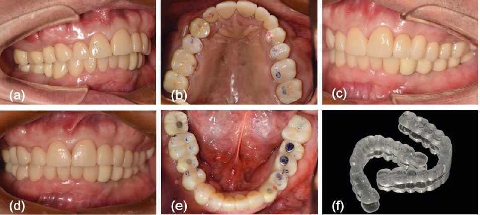
Figure 7a-fDefinitive restorations at delivery, with the protective nocturnal splint. Decision and rationale. Deliver the restorations after clinical adjustment and provide a protective splint. The report used clinical occlusal checks and instrument-assisted assessment at delivery.
Checkpoint. Judge fit and contacts independently of the EMG report.
Evidence limit. These are delivery images, not photographs dated to five-year follow-up.
Step 7 of 7 · Immediate digital occlusal assessment
Separate the check from the outcome
Previous: step 6Final step

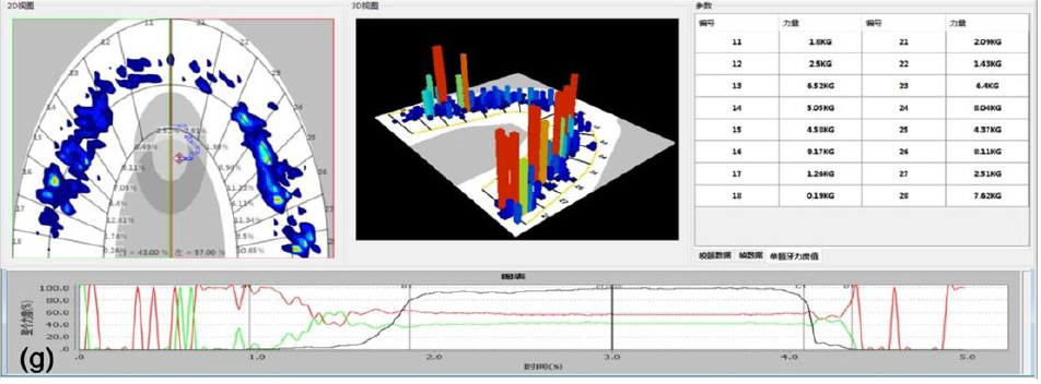
Figure 7gDigital occlusal assessment after delivery-stage adjustment. Five-year outcomes are reported separately in the article text. Decision and rationale. Keep the immediate contact assessment separate from long-term patient and restoration outcomes. The report includes a delivery-stage assessment and a later clinical maintenance history, but no dated five-year image or serial EMG dataset.
Checkpoint. Use the declared time point and distinguish each measurement from clinical benefit.
Evidence limit. The display cannot establish TMD diagnostic validity, lasting muscle relaxation, causality or comparative effectiveness.
Source: Figure 7g; section 2.3.5 and Discussion; PDF pages 6-8
What was reported over five years?
At delivery, the authors documented clinical and digital occlusal checks after adjustment (Figure 7). Separately, they reported maintained contacts and prosthesis integrity over five years, with three minor occlusal adjustments and no listed major biological or mechanical events. Satisfaction was recorded with a study-specific, unvalidated scale. No dated five-year clinical photograph or serial EMG comparison is supplied. This single case does not establish that EMG caused the outcome or validates EMG diagnosis. Source report and discussion.
What should you check when comparing EMG systems?
Start with the intended task and the evidence needed, not the number of colored report indices. Ask whether the system provides usable raw traces, documented processing, an appropriate reference protocol and validation for the interpretation you intend to make. Ease of acquisition and diagnostic accuracy are different purchasing questions.
For example, the official BioResearch BioEMG III page describes eight simultaneous channels and BioPAK displays. Cometa’s Myowise dental system describes wireless acquisition, normalized indices, raw-data access and reports. These are manufacturer-described functions, not independent evidence that either system diagnoses TMD or improves patient outcomes. Consult the exact current product documentation and safety instructions rather than assuming all dental EMG systems share the same capabilities.
Surface recording is noninvasive, but “no risk or discomfort” is too absolute. BioResearch’s BioEMG III safety notice specifies the appropriate reference electrode and reports skin-irritation events associated with malfunction and improper electrode use. The notice does not establish a complication rate; it reinforces the need for device-specific operating practice.
How should the result change the next clinical step?
For the dentist, use this evidence-led sequence:
- Establish the clinical problem. TMD symptoms and competing explanations need history and examination; an isolated amplitude or asymmetry does not replace them.
- Make the recording comparable. Correct technical problems and document task, placement and processing before interpreting a change.
- Check the claimed meaning. An event count needs validation for that device, protocol and population. A normalized index is not automatically a diagnosis.
- Measure benefit independently. Follow pain, function and the relevant dental outcome. Do not use the trace alone to justify irreversible bite changes or repeat injections.
For a technician receiving an EMG report with a restorative or appliance case, clarify what the dentist wants it to inform. Do not convert an electrical asymmetry into an unsupplied occlusal or appliance-design prescription. This is a DDS educational application of the measurement and treatment limits above; it is not evidence that EMG-guided laboratory changes improve outcomes.
Used with a clear question, surface EMG can describe muscle activity. Its clinical value depends on preserving the distinction between the signal, the interpretation and the patient’s actual outcome.
DDS connects these source findings with practical digital-dentistry decisions. Explore DDS digital-dentistry education for further learning.

WHERE DIGITAL MEETS CLINICAL
DIGITAL DENTISTRY SCHOOLOGY

Perio-Implantologist
International Digital Dentistry Speaker
Digital Occlusion Consultant
Founder of DDS
Founder of Occlusa AI Platform
 h.sharshar@ddschoology.com
Written by Dr Haitham Sharshar
Dr. Haitham Sharshar is an international digital dentistry speaker, educator, and digital occlusion consultant based in Cairo, Egypt. His work focuses on functional digital dentistry, CAD/CAM, implantology, jaw-motion analysis, and the integration of digital diagnostics into clinical treatment planning.
As Founder and Scientific Coordinator of Digital Dentistry Schoology (DDS), he has trained more than 3,270 dentists and dental technicians through university programs, international conferences, and hands-on courses. His teaching connects patient-specific diagnostic records with digital design and clinical workflows, helping clinicians and technicians understand how function and occlusion inform restorative planning.
Dr. Sharshar is a certified trainer for zebris JMA-Optic+ jaw-motion analysis and MyoWise dental EMG. His educational and consulting work brings together patient diagnostics, jaw-motion records, muscle-activity data, digital occlusion, and AI-supported workflows.
He is Founder and Clinical Director of Occlusa, an AI-supported platform for organizing clinical information and supporting clinician-led review and treatment planning. He also owns HS Dental Clinic in Cairo, where his clinical focus includes full-mouth digital rehabilitation and smile design.
His speaking and training topics include functional digital dentistry, digital occlusion, jaw-motion analysis, CAD/CAM workflows, digital implantology, and the practical integration of AI-supported tools into dental education and clinical practice.
Happy to collaborate on spreading digital dentistry.