
TMJ Disc Displacement With and Without Reduction
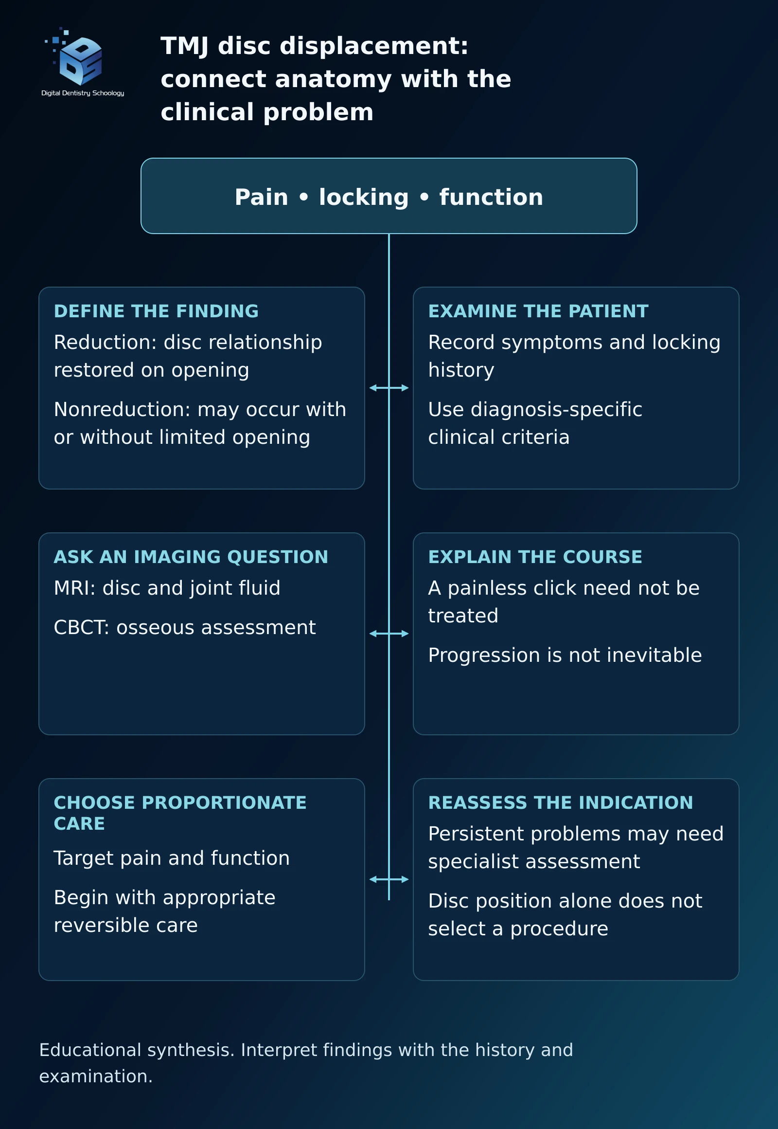
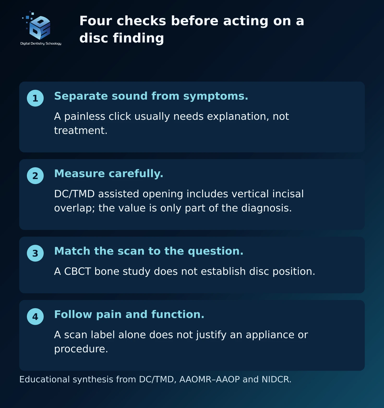
When does a clicking or locked jaw need treatment? A painless click and a jaw that suddenly will not open normally should not lead to the same conversation. Temporomandibular joint (TMJ) disc displacement describes an anatomical relationship. Treatment decisions also need the patient’s pain history, locking symptoms and functional limitations. Painless joint sounds are common and generally need no treatment; a scan label alone does not establish the cause of pain. NIDCR: temporomandibular disorders
For dentists, the useful sequence is to identify the clinical problem, decide whether imaging would answer an important question, and select care proportionate to that problem. This DDS review connects those decisions with published MRI examples, rather than treating every displaced disc as a joint that must be corrected.
What is the difference between reduction and nonreduction?
The articular disc lies between the mandibular condyle and temporal component of the joint. With disc displacement with reduction, a displaced disc regains its functional relationship with the condyle during opening. Clicking can accompany this movement. An intermittent locking history adds a different clinical feature: the jaw sometimes catches or locks with limited opening, then unlocks. DC/TMD diagnostic framework
With disc displacement without reduction, the disc remains displaced during opening. This includes two clinically distinct presentations: with limited opening and without limited opening. Nonreduction is therefore not synonymous with permanent restriction, severe pain or a mandatory surgical indication.
The DC/TMD distinction uses maximum assisted opening, including vertical incisal overlap: less than 40 mm for the limited-opening subtype and at least 40 mm for the subtype without limited opening. These measurements belong within the full diagnostic criteria, including an appropriate locking history. A single opening measurement does not establish disc displacement. Schiffman and colleagues, 2014
What should the clinical examination establish?
Begin with the complaint rather than the joint sound. Ask about pain, whether jaw movement changes it, episodes of catching or locking, and difficulty eating. Record the patient’s opening history and measured movement. Examination for familiar pain helps distinguish a pain diagnosis from an anatomical disc diagnosis; muscle and joint findings should not be collapsed into one label. DC/TMD examination and diagnostic criteria
Clinical criteria have different performance for different disc disorders. In the 2014 DC/TMD validation, the reported estimates were:
- Disc displacement with reduction
- Sensitivity 0.34 · Specificity 0.92
- With reduction and intermittent locking
- Sensitivity 0.38 · Specificity 0.98
- Without reduction, with limited opening
- Sensitivity 0.80 · Specificity 0.97
- Without reduction, without limited opening
- Sensitivity 0.54 · Specificity 0.79
These figures describe the study’s clinical criteria, not MRI accuracy. The low sensitivity for reducing displacement means that a negative clinical examination cannot confidently exclude every anatomical displacement. Conversely, an anatomical finding does not establish which structure is responsible for pain. Use the examination to frame the clinical question and recognize when diagnostic uncertainty matters. DC/TMD validity results
For the separate question of muscle-activity measurement, see DDS: dental surface EMG, its uses and interpretation limits. Surface EMG records muscle electrical activity; it does not demonstrate disc position and is not a substitute for disc-focused clinical assessment or MRI when indicated.
When does MRI add useful information?
If disc position needs imaging assessment, the AAOMR–AAOP position statement recommends closed- and open-mouth proton-density or T1-weighted MRI. T2-weighted imaging is added when effusion is suspected. CBCT evaluates osseous structures but does not directly demonstrate the articular disc or joint effusion. Choosing CBCT because it is available does not answer a soft-tissue question. Mallya and colleagues, Recommendation 4
The practical question is: What would knowing the internal-joint anatomy change? Persistent locking, restricted function or planning an intervention may make that information relevant. A painless click alone does not mean that every patient needs an MRI. This decision principle combines the imaging statement with NIDCR guidance; it is not a universal scan-ordering rule. NIDCR
What can closed- and open-mouth images show?
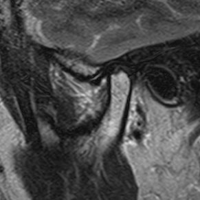
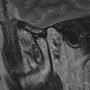
The following examples come from two different patients in a published MRI study. Each pair compares mouth position within one patient. Neither pair shows treatment or recovery.
Example 1: reduction on opening. The source describes a 56-year-old woman with bilateral disc displacement with reduction. These right-joint views are Figure 1a and 1b.
Example 2: no reduction on opening. A different patient, a 38-year-old woman, had bilateral disc displacement without reduction. These right-joint views are Figure 2a and 2b.
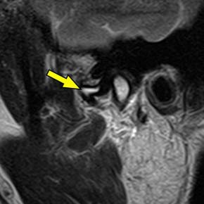
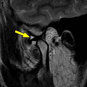
The teaching task is to compare the paired relationships and distinguish disc position from fluid. These images do not tell us how much pain either patient had or whether a particular treatment helped.
Does clicking inevitably progress to a closed lock?
No fixed sequence applies to every joint. In one small untreated cohort, one of 24 patients developed locking over an average follow-up of 25.8 months. A separate two-year study found that intermittent locking could accompany loss of disc reduction, often without permanent locking symptoms. These studies support a variable course; they do not provide a universal progression probability for an individual patient. Sato and colleagues, 2003, Kalaykova and colleagues, 2010
This distinction changes how clicking is explained. Educate the patient about reporting new pain, persistent locking or meaningful functional change, rather than presenting a painless sound as an inevitable route to joint destruction. That is a practical interpretation of the natural-history findings and NIDCR guidance.
What should initial treatment aim to improve?
The target is the patient’s pain and function, not a normal-looking disc position in every scan. NIDCR recommends starting with simple, reversible approaches when care is needed. These can include education, temporary food-texture modification, reducing aggravating habits, and appropriately selected exercises or physical therapy. Medication decisions require the individual’s medical assessment. NIDCR treatment guidance
A randomized trial of 106 people with closed lock compared medical management, rehabilitation, arthroscopy with rehabilitation, and arthroplasty with rehabilitation. Outcomes improved across strategies without a between-group advantage in the principal outcomes through 60 months. The trial supports a conservative-first discussion, but it did not include an arthrocentesis arm and should not be cited as if it did. Schiffman and colleagues, 2007
Guideline populations also matter. The 2023 BMJ guideline addresses chronic TMD pain lasting at least three months. It favors approaches including supervised exercise, mobilization and cognitive behavioral therapy, while conditionally recommending against reversible splints, arthrocentesis and botulinum toxin for that broad chronic-pain population. These recommendations should not be automatically transferred to every recent closed-lock presentation. BMJ clinical practice guideline
When should specialist procedures or appliances be discussed?
Persistent, troublesome intra-articular symptoms may justify specialist assessment. The 2024 AAOMS position paper addresses intra-articular pain and dysfunction and includes nonsurgical and minimally invasive approaches. Its scope differs from the broad chronic-pain guideline. Disc position alone should not select the procedure. AAOMS position paper
Arthrocentesis is not an automatic next step after an arbitrary number of weeks. In a separate randomized trial of 24 patients, conservative and arthrocentesis-based care produced similar pain and opening outcomes at one year. Discuss the clinical indication, alternatives and uncertainty rather than promising a predictable success rate. Öhrnell Malekzadeh and colleagues, 2019
An appliance likewise needs an explicit purpose. Do not promise that a splint will permanently reposition the disc or use an unverified fixed wear schedule for every patient. NIDCR notes limited evidence for splints in TMD pain and advises avoiding appliances intended to permanently change the bite. NIDCR
For a laboratory receiving an appliance prescription, the relevant action is clarification: identify the prescribed appliance and its intended purpose, and return unresolved design or bite-change instructions to the dentist. A scan label is not a fabrication prescription. This is a DDS educational application of the reversible-care principle, not a tested laboratory treatment protocol.
Botulinum toxin injected into masticatory muscles is not an established method for correcting disc displacement. NIDCR describes mixed TMD evidence and notes that it is not FDA-approved for TMD. A myofascial-pain trial reported dose-related adverse effects: temporarily reduced masticatory performance, reduced muscle thickness, and reduced bone volume in the mandibular coronoid and condylar processes. Evidence about muscle pain cannot be relabeled as evidence of disc repositioning. NIDCR, De la Torre Canales and colleagues, 2020
How did one symptomatic TMJ case move from assessment to digital splint delivery?
Liu and colleagues followed one 38-year-old woman with symptomatic bilateral anterior disc displacement with reduction. Their report connects pretreatment MRI, a virtual dental patient, maxillary functional anterior repositioning splint (FARS) design, fabrication and delivery. This is not a DDS patient, and it is separate from the two Mizuhashi MRI examples above.
This is a documented single-case workflow, not a general appliance recommendation. Five intact source figures show the assessment, digital stages and delivered appliance. The sixth step reports the three-month outcome as text: no reliably dated follow-up image is presented, so visual outcome coverage is incomplete. The case does not override the management guidance above.
Use the numbered links or previous/next links, or swipe the figures sideways. Check “Read all steps in one vertical list” to read down the page; uncheck it to return to slides. Open a full figure to inspect small labels. All captions and explanations remain available without JavaScript.
Step 1 of 6 · Clinical assessment · pretreatment MRI
Establish the clinical and MRI baseline
First stepNext: step 2

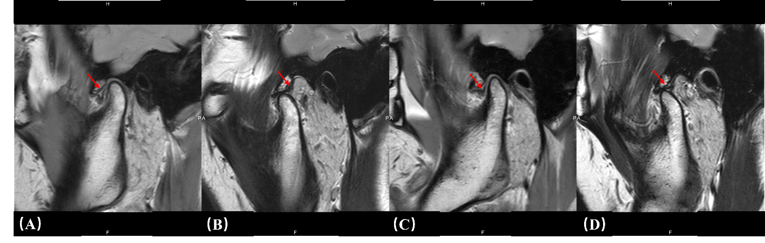
Pretreatment MRI: left TMJ in intercuspation (A) and maximum opening (B), then right TMJ in intercuspation (C) and maximum opening (D). The authors describe bilateral anterior disc displacement that reduces on opening. Liu et al., Supplementary Figure S2. Open full Supplementary Figure S2
Decision and rationale. Authors established bilateral ADDwR before splint planning. MRI and clinical assessment provide the case diagnosis; the motion graphs are not substitutes for imaging of the disc.
Checkpoint and limitation. Read the paired closed/open positions in source order: left joint A–B, right joint C–D. These publication panels are not the complete MRI examination; DDS is not independently diagnosing the patient from them.
Step 2 of 6 · Digital records · virtual patient
Combine records in a virtual patient

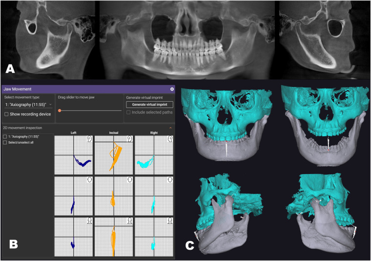
The reported virtual patient combines intraoral scans, CBCT-derived jaw models and Modjaw movement records: CBCT views (A), motion traces (B), and the fused model (C). Liu et al., Figure 1. Decision and rationale. Authors combined geometry and jaw-motion records in exocad. The fused record enabled the authors to review dynamic tooth contacts and mandibular positions.
Checkpoint and limitation. The tracker in this case was Modjaw. The surface scans, CBCT and motion records do not directly image the articular disc or establish disc recapture.
Step 3 of 6 · Digital planning · bone relationships
Select a mandibular position for planning

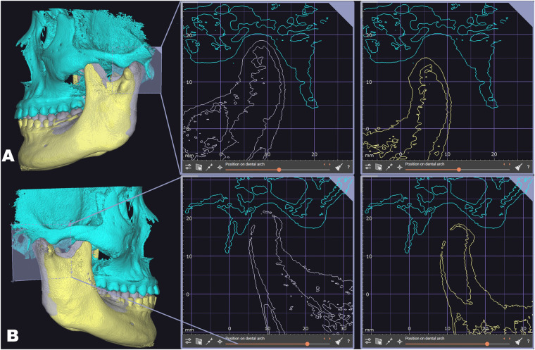
The authors compared the initial mandibular position (gray) with their selected forward position (yellow), at the left joint (A) and right joint (B). These are bone-position planning views, not direct evidence of disc recapture. Liu et al., Figure 2. Decision and rationale. Authors selected an anterior mandibular position using the recorded movement and virtual bone relationships. The source describes a proposed position for anterior repositioning splint design.
Checkpoint and limitation. Gray is the initial position and yellow the selected forward position. This figure documents a planning choice, not MRI proof of disc recapture or a numerical protrusion target for other patients.
Step 4 of 6 · Digital design · CAD
Design the maxillary splint

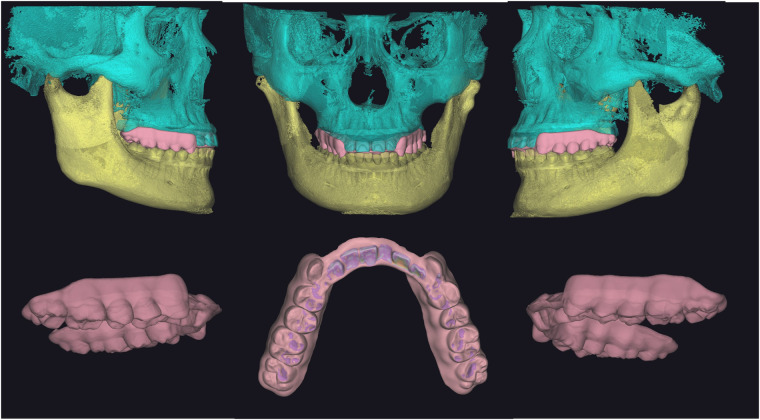
The maxillary FARS was designed with patient-specific occlusal morphology and an anterior palatal guard. The approximately 2 mm thickness is this case’s reported design, not a general prescription. Liu et al., Figure 3. Decision and rationale. Authors tailored the splint surface and anterior guard to their selected relation and recorded dynamic occlusion. The reported goals were stable contact, lateral guidance and resistance to posterior mandibular return.
Checkpoint and limitation. The upper-arch CAD design is not evidence of manufactured fit, accuracy or clinical safety. Its reported approximately 2 mm thickness is case-specific.
Step 5 of 6 · Manufactured appliance · clinical delivery
Inspect the fabricated appliance and its delivery

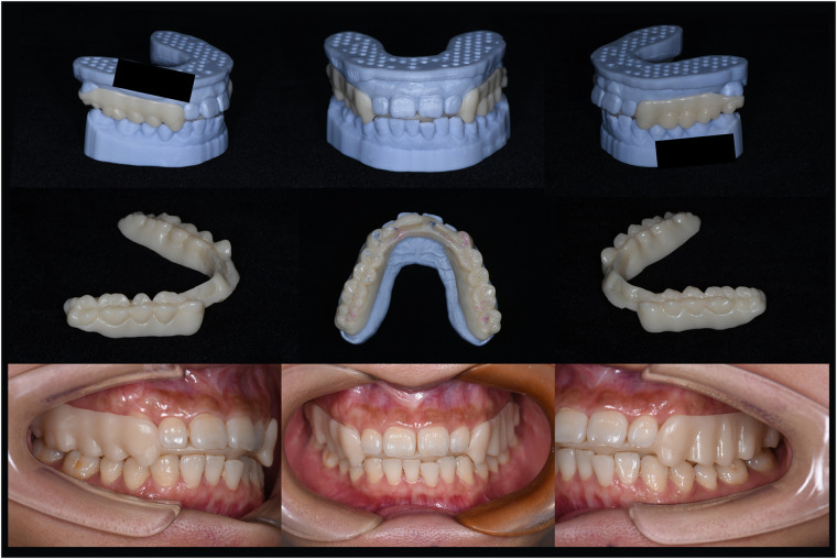
Top and middle rows: the fabricated splint, alone and on dental casts. Bottom row: three intraoral views at delivery; these are not the three-month follow-up. The whole published Figure 4 is retained, including the original black masks. It shows a manufactured result and delivery, not the printing process. Liu et al., Figure 4. Fabrication decision. The authors report a Chairside Pro printer and Surgical Guide UV resin. The physical appliance is distinct from the preceding CAD design, but the paper supplies no reproducible printing, washing or post-curing protocol.
Delivery decision and rationale. The authors assessed the seated splint during oral movements and chewing, bringing the planned appliance into a clinical check. Static photographs cannot verify dynamic performance.
Checkpoint and limitation. The material and device names describe what the authors reported; they are not a DDS recommendation for manufacture or prolonged wear. Exact material-version and processing suitability for the reported use are not established here. These official product links identify current products; they are not a validated long-wear instructions-for-use (IFU) protocol or evidence for the case outcome.
Step 6 of 6 · Clinical follow-up · source-reported text
Reported three-month outcome (text only)
Previous: step 5Final step
Source-reported outcome. At three months, the patient reported substantially less jaw pain and clicking, and the authors described comfortable chewing. Orthodontic treatment was still ongoing. This single case does not establish durable disc recapture or comparative effectiveness.
Decision and rationale. Read the report’s symptom and chewing observations as a short follow-up of this one patient. They do not isolate the appliance’s causal effect or establish superiority over other care.
Evidence boundary. No dated follow-up MRI is shown. Supplementary Figure S3 has inconsistent timing between its caption and the outcome text, so it is excluded from this sequence. There is no invented outcome photograph, percentage improvement or claim that treatment was finished.
What should the dentist take back to practice?
- Name the problem before treating the anatomy. Record pain, locking and function; apply diagnosis-specific criteria rather than interpreting every click as disease.
- Match the image to the question. Use MRI when internal-joint information is needed; a CBCT bone assessment does not establish disc position.
- Choose outcomes the patient can recognize. Follow pain and meaningful function separately from the scan. Begin with proportionate, reversible care and reassess persistent problems.
- Keep the prescription specific. Specialist procedures and appliances need a clinical indication, an explanation of uncertainty and follow-up. Neither the MRI label nor a generic treatment ladder supplies that reasoning.
These are educational conclusions from the linked diagnostic, imaging and management sources. They support clinical reasoning; they do not replace an individual examination.
DDS connects these source findings with practical digital-dentistry decisions. Explore DDS digital-dentistry education for further learning.

WHERE DIGITAL MEETS CLINICAL
DIGITAL DENTISTRY SCHOOLOGY

Perio-Implantologist
International Digital Dentistry Speaker
Digital Occlusion Consultant
Founder of DDS
Founder of Occlusa AI Platform
 h.sharshar@ddschoology.com
Written by Dr Haitham Sharshar
Dr. Haitham Sharshar is an international digital dentistry speaker, educator, and digital occlusion consultant based in Cairo, Egypt. His work focuses on functional digital dentistry, CAD/CAM, implantology, jaw-motion analysis, and the integration of digital diagnostics into clinical treatment planning.
As Founder and Scientific Coordinator of Digital Dentistry Schoology (DDS), he has trained more than 3,270 dentists and dental technicians through university programs, international conferences, and hands-on courses. His teaching connects patient-specific diagnostic records with digital design and clinical workflows, helping clinicians and technicians understand how function and occlusion inform restorative planning.
Dr. Sharshar is a certified trainer for zebris JMA-Optic+ jaw-motion analysis and MyoWise dental EMG. His educational and consulting work brings together patient diagnostics, jaw-motion records, muscle-activity data, digital occlusion, and AI-supported workflows.
He is Founder and Clinical Director of Occlusa, an AI-supported platform for organizing clinical information and supporting clinician-led review and treatment planning. He also owns HS Dental Clinic in Cairo, where his clinical focus includes full-mouth digital rehabilitation and smile design.
His speaking and training topics include functional digital dentistry, digital occlusion, jaw-motion analysis, CAD/CAM workflows, digital implantology, and the practical integration of AI-supported tools into dental education and clinical practice.
Happy to collaborate on spreading digital dentistry.