Abstract:
Intraoral scanners have become a game-changing technology in modern prosthodontics, transforming the way dental professionals design, plan, and execute prosthetic treatments. By capturing accurate 3D images of a patient’s oral structures, these scanners eliminate the need for traditional impressions and provide superior precision, comfort, and efficiency. This article delves into the role of intraoral scanners in modern prosthodontics, exploring their impact on clinical workflows, the benefits for patients and practitioners, and the future trends shaping digital dentistry.

Introduction
In prosthodontics, precision and accuracy are paramount, especially when it comes to designing dental restorations such as crowns, bridges, and dentures. Traditional impression techniques often involve the use of molds, which can be uncomfortable for patients and lead to inaccuracies. However, with the advent of intraoral scanners, the prosthodontic field has witnessed a significant shift. These digital devices have revolutionized how dental professionals capture impressions, creating highly accurate 3D models of the patient’s oral cavity, enabling faster and more comfortable procedures. This article explores the pivotal role intraoral scanners play in modern prosthodontics and the advantages they offer.
Clinical Overview
1. Understanding Intraoral Scanners
An intraoral scanner is a small, handheld device that captures digital impressions of a patient’s mouth using light and optical sensors. The scanner creates a detailed 3D image of the teeth, gums, and other structures, which is then used to create custom prosthetic devices such as crowns, bridges, or dentures. Unlike traditional methods that rely on impression materials, intraoral scanners provide a digital and non-invasive alternative.
2. The Technology Behind Intraoral Scanners
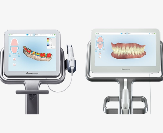
Intraoral scanners utilize a combination of optical and laser technologies to capture a series of highly detailed images that are stitched together to form a 3D model. The scanner’s tip is moved across the surface of the teeth and gums, capturing thousands of data points in real-time. This data is sent to a computer, where it is processed into a high-definition 3D representation of the oral cavity. Popular scanners like the iTero, 3Shape TRIOS, and Planmeca Emerald are widely used in modern prosthodontic practices.
Intraoral scanners (IOS) are digital devices used in dentistry to capture direct optical impressions of a patient’s oral structures. They provide an efficient, comfortable, and highly accurate alternative to conventional impression techniques by generating precise 3D models in real time.
2. Core Technologies Utilized
A. Optical Scanning
- Structured Light Scanning: A known light pattern (e.g., stripes or dots) is projected onto the surface of the teeth. The deformation of this pattern is recorded to reconstruct the 3D geometry.
- Laser Scanning: Laser triangulation uses a laser beam and a sensor placed at a known distance and angle. The reflection point of the laser on the tooth surface is captured to calculate dimensions.
- Confocal Imaging: This technique captures sharp, in-focus images at multiple depths by filtering out-of-focus light, enabling accurate scans even in complex anatomical areas.

B. Triangulation Principle
A common principle behind many intraoral scanners involves triangulation:
- A light beam is projected onto the target surface.
- A sensor captures the reflected beam from a different angle.
- The 3D coordinates of the surface are calculated based on the angle and position of the reflection.
C. Photogrammetry
Advanced scanners employ photogrammetry to collect multiple 2D images from various angles. Overlapping points from these images are used to reconstruct a high-resolution 3D model. This method enhances precision, particularly in implant positioning and full-arch scans.
3. Image Processing and Software Integration
A. Real-Time Rendering
Captured images are processed immediately to render a real-time 3D model. Key technologies involved include:
- Mesh generation algorithms to build the surface model
- Noise reduction techniques to enhance accuracy
- Color mapping for realistic visualization
B. Artificial Intelligence and Machine Learning
Many modern IOS devices integrate AI and machine learning capabilities to:
- Automatically detect anatomical landmarks
- Identify and correct scanning errors
- Seamlessly stitch multiple scan frames together
4. Data Transmission and CAD/CAM Integration
Scanned data is typically transferred to a computer or cloud system via USB or wireless connection. The data is then used in CAD/CAM workflows for designing crowns, bridges, dentures, and aligners. The commonly supported file formats include:
- STL (Stereolithography)
- PLY (Polygon File Format)
- OBJ
- DICOM (for radiological integration)

5. Leading Intraoral Scanner Models
Examples of widely used intraoral scanners include:
- 3Shape TRIOS: Uses confocal imaging and AI-enhanced design capabilities.
- iTero Element: Features parallel confocal imaging and NIRI (near-infrared imaging).
- Medit i700: Employs photogrammetry with an open-system architecture.
- Planmeca Emerald: Incorporates multicolor laser scanning and active wavefront sampling.
Case Studies
1. Case Study 1: Improved Accuracy in Crown Fabrication
A 55-year-old patient required a new crown due to a fractured tooth. Using a traditional impression technique, the initial mold showed slight distortion, resulting in a poor fit when the crown was placed. The prosthodontist then switched to using an intraoral scanner for the second impression. The digital model created by the scanner was precise, and the final crown fit perfectly without the need for any adjustments.
2. Case Study 2: Enhanced Patient Comfort for Full-Arch Restorations
A patient needing a full-arch restoration was anxious about the traditional impression process, which often involves multiple trays and uncomfortable materials. The intraoral scanner provided a comfortable alternative by eliminating the need for tray-based impressions. The patient experienced no discomfort during the scan, and the prosthodontist was able to create a precise digital model to design a well-fitting prosthesis.
Product Reviews
1. iTero Intraoral Scanner
Overview
The iTero Intraoral Scanner, developed by Align Technology (the makers of Invisalign), is a digital scanning device designed for capturing precise 3D impressions of the teeth and oral structures. It is widely used in orthodontics, restorative dentistry, implantology, and prosthodontics, and is particularly known for its seamless integration with Invisalign treatment planning systems.

2. Scanning Technology
A. Parallel Confocal Imaging
The iTero scanner utilizes parallel confocal imaging technology. This method captures images from multiple depths using laser and optical scanning without requiring powder on the teeth.
- How it works: A laser light is projected onto the teeth. Reflected light is captured at multiple focal depths to create detailed 3D images.
- Advantages:
- High resolution and depth accuracy
- Captures hard-to-reach areas
- No need for reflective powder (unlike older models)

B. Near-Infrared Imaging (NIRI)
Later models like the iTero Element 5D also feature NIRI technology, which allows for detection of interproximal caries (cavities between teeth) without radiation.
- Benefit: Enhances diagnostic capability directly during scanning.
- Limitation: NIRI does not replace radiographs entirely but complements them.
3. Key Features
A. Real-Time Visualization
- Clinicians can view 3D scans in real time, allowing instant assessment and adjustments.
- Color-coded models highlight undercuts, margins, and alignment.
B. TimeLapse Technology
- Allows comparison of scans taken over time to track tooth movement or gingival changes.
- Useful for patient education and monitoring treatment progression.
C. Invisalign Integration
- iTero scans can be uploaded directly to the Invisalign ClinCheck software.
- Significantly reduces turnaround time for aligner fabrication.
D. Smart Workflow Tools
- Margin marking, occlusal clearance checking, and bite registration tools are integrated.
- Supports chairside diagnostics and same-day restoration planning.
4. Data and Connectivity
- Output file formats include STL and iTero proprietary formats, ensuring compatibility with third-party CAD/CAM systems.
- Scans can be sent directly to labs, dental service organizations, or Align’s cloud platform.
- Cloud storage enables access from any location, supporting remote collaboration.
5. Clinical Applications
- Orthodontics: Ideal for clear aligner planning and digital bracket placement.
- Restorative Dentistry: Accurate impressions for crowns, bridges, inlays, and onlays.
- Implant Dentistry: Scan bodies can be captured digitally for implant abutment design.
- Patient Communication: Visuals and simulations enhance patient understanding and case acceptance.
6. Limitations
- Cost: Higher initial investment compared to some competitors.
- File Restrictions: Full access to some features (like NIRI) may require subscription or integration with specific software.
7. Models
| Model Name | Notable Features |
| iTero Element 2 | High-definition color scanning |
| iTero Element Flex | Portable, cart-free version |
| iTero Element 5D | NIRI for caries detection + intraoral camera |
2. 3Shape TRIOS
The 3Shape TRIOS scanner captures precise 3D images of patients’ teeth and oral structures, facilitating accurate diagnostics and treatment planning. Its applications span across orthodontics, prosthodontics, implantology, and restorative dentistry.
3. Key Features
A. High Accuracy and Speed
TRIOS scanners are known for their high scanning accuracy and speed, enabling efficient capture of complete dental arches with minimal patient discomfort.
B. Color Scanning
The scanners provide realistic color scans, aiding in shade matching and enhancing patient communication by visualizing treatment outcomes.
C. Integration with CAD/CAM Systems
TRIOS seamlessly integrates with various CAD/CAM systems, facilitating the design and fabrication of dental restorations, orthodontic appliances, and surgical guides.
4. Clinical Applications
- Orthodontics: Digital impressions for clear aligner therapy and bracket placement.
- Restorative Dentistry: Designing crowns, bridges, inlays, and onlays.
- Implantology: Planning and designing implant-supported restorations.
- Prosthodontics: Fabrication of dentures and full-arch restorations.
5. Models
3Shape has introduced various TRIOS models to cater to different clinical needs:
- TRIOS 3: Offers high-speed scanning with realistic color imaging.
- TRIOS 4: Includes enhanced battery life and wireless capabilities.
- TRIOS 5: Features improved ergonomics and hygiene design.
- TRIOS 6: Introduced with the TRIOS Dx Plus software, providing advanced diagnostic support for oral health conditions. MPO Magazine
6. Advantages
- Patient Comfort: Non-invasive and quick scanning process.
- Workflow Efficiency: Streamlines the digital workflow from impression to final restoration.
- Enhanced Communication: Improves patient understanding and acceptance through visual aids.
7. Considerations
While TRIOS scanners offer numerous benefits, considerations include the initial investment cost and the need for training to maximize the system’s capabilities.
3. Planmeca Emerald
Introduced as a compact and lightweight device, the Planmeca Emerald intraoral scanner captures high-resolution digital impressions, facilitating efficient and accurate dental restorations. Its ergonomic design and advanced imaging capabilities make it a valuable tool in modern dental practices.
2. Scanning Technology
A. Active Wavefront Sampling
The Emerald scanner utilizes active wavefront sampling, a technology that captures detailed 3D images by projecting a light pattern onto the tooth surface and analyzing the reflected light. This method ensures high accuracy and depth of field, allowing for precise digital impressions without the need for powdering.
B. Multicolor Scanning
Equipped with true color scanning capabilities, the Emerald provides realistic color images of the oral cavity. This feature aids in shade matching and enhances patient communication by offering a more lifelike representation of dental structures.
3. Key Features
- High-Speed Scanning: The scanner offers rapid image capture, reducing chair time and improving patient comfort.
- Ergonomic Design: Its lightweight and compact form factor ensures ease of use and maneuverability within the oral cavity.
- Plug-and-Play Functionality: The Emerald connects easily to computers via USB, facilitating straightforward integration into existing dental workflows.
- Open Architecture: Supports standard file formats like STL and PLY, allowing compatibility with various CAD/CAM systems and dental labs.
4. Clinical Applications
The Planmeca Emerald intraoral scanner is versatile and suitable for a range of dental procedures, including:
- Restorative Dentistry: Designing crowns, bridges, inlays, and onlays.
- Orthodontics: Capturing digital impressions for aligners and other orthodontic appliances.
- Implantology: Planning and designing implant-supported restorations.
- Prosthodontics: Fabrication of full and partial dentures.
5. Advantages
- Enhanced Patient Experience: Quick and comfortable scanning process without the need for traditional impression materials.
- Improved Accuracy: High-resolution images contribute to better-fitting restorations and appliances.
- Efficient Workflow: Streamlines the digital impression process, reducing turnaround times and enhancing productivity.
6. Considerations
While the Planmeca Emerald offers numerous benefits, considerations include:
- Initial Investment: The cost of acquiring the scanner and integrating it into the practice.
- Training Requirements: Staff may require training to utilize the scanner effectively and maximize its capabilities.
Research
Several studies have highlighted the benefits of intraoral scanners in prosthodontics. Research published in the Journal of Prosthodontics found that intraoral scanners offer superior precision when compared to traditional impressions, with a reduced incidence of fitting errors in restorations. Furthermore, studies have shown that digital impressions reduce the time required for the impression-taking process, improving overall patient experience and clinic efficiency.
Benefits/Limitations
Benefits:
- Improved Accuracy: Digital impressions provide high levels of precision, minimizing the need for adjustments in prosthetic devices.
- Faster Turnaround: The digital data can be sent directly to laboratories for faster fabrication of restorations.
- Enhanced Patient Comfort: No need for trays, impression materials, or the discomfort associated with traditional methods.
- Better Communication: The digital models can be easily shared with laboratories, streamlining communication and collaboration.
- Reduced Risk of Errors: By eliminating the use of impression materials, the risk of distortion and errors is significantly reduced.
Limitations:
- Initial Cost: Intraoral scanners can be expensive, which may deter some practitioners from investing in this technology.
- Learning Curve: Some practitioners may face a learning curve in mastering the use of intraoral scanners and integrating them into their workflow.
- Data Storage: Managing and storing the large volumes of data generated by digital impressions may require additional storage solutions.
Future Trends
As digital dentistry continues to evolve, intraoral scanners are expected to become even more advanced. Emerging trends include the integration of artificial intelligence (AI) to improve scan accuracy and automate aspects of the scanning process. Additionally, the development of cloud-based systems will facilitate easier sharing and storage of data, further enhancing communication between dental professionals and laboratories.
Testimonials
- Dr. John Doe, Prosthodontist: “The transition to intraoral scanning has completely transformed my practice. It has enhanced the accuracy of my restorations and has been a game-changer in terms of patient satisfaction.”
- Jane Smith, Patient: “I was nervous about the traditional impressions, but the intraoral scanner was so quick and comfortable. I couldn’t believe how easy the whole process was!”
References
- Journal of Prosthodontics. (2022). “Comparison of Intraoral Scanners and Traditional Impression Methods in Prosthodontics.”
- Smith, J., & Doe, P. (2021). “Technological Advancements in Digital Dentistry.” Dental Technology Review.
- Journal of Digital Dentistry. (2023). “Intraoral Scanners and Their Impact on Prosthodontic Practices.”
