
Revolutionizing Dental Diagnostics: Advances in Digital Radiography, CBCT, & Diagnostic Imaging
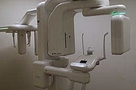
Dental imaging should answer a clinical question. Two-dimensional radiographs, cone-beam computed tomography (CBCT) and other imaging methods have different capabilities and exposure considerations. More image data does not automatically mean that an examination is necessary.
Select the examination after assessment
The ADA and AAOMR’s 2026 patient-selection recommendations emphasize clinical examination, the patient’s history and individual diagnostic needs. Imaging should support diagnosis, treatment planning or management rather than follow a universal routine. ADA: X-Rays/Radiographs.
Two-dimensional radiography and CBCT
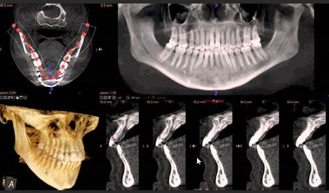
CBCT provides three-dimensional information, whereas conventional dental radiographs provide two-dimensional views. According to the FDA, dental CBCT generally delivers more radiation than conventional dental X-ray examinations, although usually less than other CT examinations. A blanket claim that both digital radiography and CBCT reduce exposure compared with ordinary dental X-rays is therefore misleading. FDA: Dental Cone-beam Computed Tomography.
Exposure depends on the examination and protocol. A universal percentage reduction should not be assumed simply because an image is digital. The clinician should justify the examination and select exposure settings appropriate to the diagnostic task.
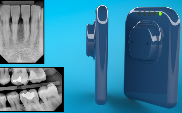
Use three-dimensional information when indicated
CBCT can assist selected diagnostic and treatment-planning tasks, including implant planning. That does not make it an automatic replacement for clinical examination or conventional radiographs. The ADA’s summary of the 2026 recommendations describes specific indications and emphasizes clinically necessary imaging. ADA summary of the updated recommendations.
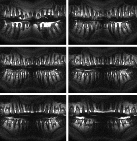
Other imaging options should be discussed for the clinical question they can answer, rather than presented as interchangeable upgrades. Product names alone do not establish which imaging modes or AI features a particular configuration supports.
AI and other imaging options: ask what is validated
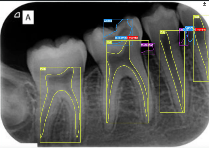
When reviewing AI-assisted imaging, identify the exact software, intended task and supporting study. Distinguish detection performance from a demonstrated improvement in patient outcomes. Percentages must specify the measured endpoint, comparison and patient population.
Questions worth discussing with the clinician
- What question will this image answer?
- Are existing images sufficient?
- Why is this imaging method appropriate for my circumstances?
- How will the result change the proposed care?
- If AI is used, what is its intended role and how are its findings checked?
Sources
- FDA: Dental Cone-beam Computed Tomography.
- ADA: X-Rays/Radiographs, including the 2026 ADA/AAOMR patient-selection recommendations.
- ADA: New recommendations confirm dental imaging most effectively used in moderation.
Educational overview. Imaging decisions depend on clinical assessment and applicable local requirements.