
Peri-Implantitis: Prevention and Management Strategies
Start with the disease status and the clinical goal
What needs to change when inflammation develops around an implant? First establish whether the problem is peri-implant mucositis or peri-implantitis, identify factors that can be addressed, and define how treatment response will be assessed. An implant remaining in place is a different outcome from resolution of disease. This overview connects diagnosis, prevention, treatment and continuing care.
Distinguish health, mucositis and peri-implantitis
- Peri-implant health: no clinical inflammation, bleeding or suppuration on gentle probing, with no increase in probing depth or bone loss beyond initial remodeling. Health can exist with reduced bone support.
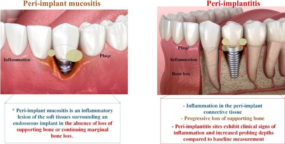
- Peri-implant mucositis: bleeding and/or suppuration on gentle probing, with or without increased probing depth, but without bone loss beyond initial remodeling.
- Peri-implantitis: inflammation with bleeding and/or suppuration, increased probing depth relative to earlier examinations, and bone loss beyond initial remodeling.
When previous examination data are unavailable, the World Workshop describes a combined diagnostic fallback: bleeding and/or suppuration, probing depth at least 6 mm, and bone level at least 3 mm apical to the most coronal intraosseous part of the implant. This applies to missing baseline records; it is not a rule specifically for newly placed implants. Recession also matters when interpreting probing depth. [1]
Read prevalence as a population estimate
Derks and Tomasi pooled a peri-implantitis prevalence of approximately 22% (95% confidence interval 14–30%) across studies with differing definitions and follow-up. That figure does not mean that 22% of all implant patients will develop disease within a specified five-to-ten-year period. A prevalence estimate describes the sampled populations; it is not an individual prognosis. [4]
Look for risks and practical barriers to care
The 2025 AO/AAP consensus identifies relevant patient-level factors including a history of periodontitis, smoking, uncontrolled diabetes and inadequate biofilm control. Local considerations include implant position, prosthetic design and peri-implant soft-tissue characteristics. These factors need interpretation together rather than conversion into an unsupported universal risk multiplier. [3]
- Before treatment: review periodontal and medical history, current tissue findings and the patient’s ability to perform home care.
- At the implant and restoration: assess position, contour and access for cleaning; identify local problems that may need correction.
- With the patient: discuss modifiable risks, treatment choices and the ongoing maintenance commitment.
The practical aim is a patient-specific plan that addresses both disease and the conditions that make it difficult to control. Correcting a prosthetic access problem and treating an inflammatory lesion are connected tasks, not interchangeable outcomes. [3]
Manage mucositis with biofilm control and reassessment
For mucositis, professional mechanical plaque removal and patient-performed oral hygiene are central. The 2015 prevention consensus reported improvement in inflammatory signs with oral-hygiene instruction and mechanical debridement; its reviewed adjuncts did not establish additional benefit over professional plaque removal. Review the tissue response and persistent contributing factors rather than assuming that a rinse or device has resolved the condition. [7]
Use a staged peri-implantitis pathway
The 2023 EFP guideline starts with nonsurgical treatment and reassessment. Persistent disease may require surgery; an isolated 2 mm bone-loss cutoff does not select the treatment. Adequate home care, prosthetic access and appropriate surgical expertise matter. [2]
Antimicrobials: routine systemic antibiotics are not recommended as nonsurgical adjuncts. Systemic antibiotics are not recommended as adjuncts to peri-implantitis surgery; evidence is insufficient for surgical local antibiotics. These are context-specific recommendations, not a ban on antibiotics for every clinical indication.
Adjuncts: for nonsurgical peri-implantitis treatment, the guideline suggests against local antimicrobials, air polishing, lasers and photodynamic therapy in their evaluated roles. During surgery, it suggests against air polishing, Er:YAG laser, chlorhexidine and photodynamic surface decontamination. Titanium brushes may be considered; there is insufficient evidence for a recommendation on implantoplasty.
Reassessment: the nonsurgical endpoint includes residual probing depths no greater than 5 mm, bleeding at no more than one site, and no suppuration. Findings and their course inform escalation. These recommendations should not be transferred indiscriminately to mucositis, maintenance or every new device. [2]
Keep follow-up evidence separate from a success story
In Heitz-Mayfield and colleagues’ study, 24 patients with 36 affected implants received an anti-infective surgical protocol followed by supportive therapy. Study-defined success was reported in 19 of 24 patients at one year and 15 of 24 at five years. Some patients experienced recurrence or implant loss, and some were lost to follow-up. This is a published cohort with defined outcomes, not proof that one adjunct caused recovery or a prediction for a new patient. It shows why favorable early findings need to be followed over time. [5]
Plan continuing care as part of treatment
The ITI consensus connects individualized supportive care with monitoring, professional and home biofilm control, and management of modifiable risks. Recurrence can occur despite maintenance. New bleeding or suppuration together with increasing probing depth warrants reassessment; radiography may help when the diagnosis is unclear. [6]
- Record the outcome: distinguish implant survival, tissue inflammation, bone-level stability and the patient’s comfort and ability to clean.
- Compare over time: retain clinical measurements and appropriate radiographic records rather than relying on a single photograph.
- Plan the next review: tailor continuing care to the patient and the findings; further treatment or implant removal may be needed if disease progresses.
These are continuing-care principles. They do not establish a universally successful surgical technique or guarantee that a treated implant will remain healthy. [6]
Questions for the dentist and laboratory
For the dentist and laboratory, check:
- Which records establish the diagnosis, and which baseline records are missing?
- Can the patient and clinician access the restoration for cleaning and assessment?
- Which proposed step addresses inflammation, which addresses a prosthetic problem, and how will each be checked?
- Does a claimed result refer to probing findings, radiographic change, implant survival or patient experience?
- What is the review plan if inflammation persists or returns?
Digital models, surface technologies and proposed regenerative adjuncts should be evaluated against a defined task and a traceable clinical source. A diagram or proposed mechanism cannot substitute for a documented outcome. The useful connection is from the patient’s findings to the treatment objective, then to verifiable follow-up.
References
- Berglundh and colleagues. Peri-implant diseases and conditions: 2017 World Workshop consensus (2018).
- Herrera and colleagues. Prevention and treatment of peri-implant diseases: EFP S3 guideline (2023).
- Wang and colleagues. AO/AAP consensus on prevention and management of peri-implant diseases and conditions (2025).
- Derks and Tomasi. Peri-implant health and disease: systematic review of epidemiology (2015).
- Heitz-Mayfield and colleagues. Supportive peri-implant therapy following anti-infective surgery: five-year survival and success (2018).
- ITI Consensus 2018. Outcomes of peri-implantitis treatment followed by supportive care.
- Jepsen and colleagues. Primary prevention of peri-implantitis: managing peri-implant mucositis (2015).