
Soft Tissue Management Around Implants: From Risk Assessment to Maintenance
Soft tissue management around implants brings together site assessment, implant positioning, augmentation, provisional contouring and long-term review. The useful clinical question is which tissue problem needs treatment: insufficient volume, limited keratinized mucosa, an unfavourable margin, or inadequate support from the underlying anatomy. These problems do not have one interchangeable graft or a fixed prosthetic solution.
This educational guide retains a staged clinical workflow while separating published observations, manufacturer guidance and planning considerations. It is not a patient-specific surgical prescription or a promise of an aesthetic result.
1. Define the tissue problem before selecting a procedure
Record the smile line, mucosal margin and thickness, keratinized mucosa, adjacent tooth support, facial bone anatomy, restorative space and ability to clean the proposed restoration. Discuss expectations and the possibility of additional treatment. Implant survival alone does not describe the appearance or health of the surrounding tissues.
Assessment should distinguish the soft-tissue envelope from its bony support. A thin phenotype or visible contour deficiency warrants closer planning; a thickness label alone should not automatically determine a submerged protocol or a graft. Similarly, a single generic set of implant distances cannot replace anatomy- and restoration-led planning.
Keratinized Tissue: In a 10-year study of posterior mandibular implants, absence of keratinized mucosa was associated with more plaque accumulation and soft-tissue recession. The study compared tissue presence with absence rather than a 2-mm cutoff, so it does not establish a universal minimum width (Roccuzzo et al., 2016).
The clinical implication is to assess tissue width alongside inflammation, plaque control, brushing comfort and recession rather than treat a numerical threshold as an isolated indication for surgery. Document baseline findings so later changes can be interpreted.
2. Plan placement timing and soft-tissue access together
Use the intended restoration to plan implant position, then check whether the site can support that position. A surgical guide or digital plan helps communicate the intended geometry; it does not establish that the bone, soft tissue or primary stability will be adequate at surgery.
The 2023 ITI selection consensus for immediate placement with immediate loading in the maxillary aesthetic zone calls for patient- and site-specific assessment and contingency planning. Extraction-related events or inadequate primary stability may prevent completion of the intended immediate protocol. Explain an alternative timing/restoration plan before surgery rather than relying on one insertion-torque cutoff. [4]
Vertical Stability: A systematic review found variable midfacial mucosal recession after immediate implant placement in the anterior maxilla; heterogeneity prevented a meta-analysis of controlled studies (Chen & Buser, 2014).
Flapless access, a limited flap and wider exposure are different ways to obtain access. Select the approach needed to assess and manage the actual defect while protecting available tissue; minimal access is not an end in itself. Papilla preservation, careful handling and suitable wound closure remain planning priorities, but no incision design guarantees freedom from recession.
3. Match graft selection to the treatment objective
Autogenous connective tissue and substitute matrices belong in a discussion of the intended gain, donor-site burden, defect anatomy, healing requirements and the evidence for that indication. A matrix that supports keratinized-tissue management is not automatically a volume-stable substitute for every connective-tissue graft. Product names should not be used as shorthand for equivalent clinical performance.
Mucograft and Fibro-Gide have different material and healing profiles
Geistlich’s professional guidance describes Mucograft as a non-cross-linked collagen matrix with reduced volume stability and options for open or submerged healing. Its listed applications include keratinized-tissue gain, vestibuloplasty, recession coverage and socket-seal treatment. Fibro-Gide is a cross-linked, volume-stable collagen matrix intended for soft-tissue volume augmentation and recession defects, with submerged healing. These are manufacturer-described distinctions, not independent proof of superior long-term aesthetics. [5]
For Fibro-Gide, the manufacturer emphasizes full coverage, tension-free closure and avoidance of compression at the defect. For either material, check the current locally applicable instructions for use, exact product variant, contraindications and handling requirements. Do not transfer the healing approach of one matrix to another. [5]
A connective-tissue graft, a roll-type approach using local tissue, or a substitute matrix may be considered for different defects. Choose a technique on the basis of the needed tissue change and the clinician’s training. The discussion should include donor-site morbidity where relevant, incomplete correction, wound complications and the possibility of further treatment, rather than an assurance of a predictable volume gain.

Keep socket preservation and soft-tissue augmentation conceptually separate
After extraction, assess socket-wall integrity and the planned implant timing before choosing a preservation or reconstruction approach. Bone-substitute placement, a barrier membrane, soft-tissue coverage and a provisional pontic perform different jobs. A named bone graft does not by itself establish the final mucosal contour or remove the need for reassessment.
4. Develop the emergence profile within tissue tolerance
The provisional restoration can communicate and test the intended contour. Distinguish restorative shape changes from implant loading, and avoid treating progressive contouring as a fixed compression prescription. Su and colleagues’ critical/subcritical contour framework describes where a change is made and what it may influence. [2]
- Subcritical contours: Located apical to the critical contour; in Su et al.’s clinical observations, modifications within tissue tolerance could change the soft-tissue profile without a clinically significant change in facial gingival margin level.
- Critical contours: Located immediately below the gingival margin; facial contour influences margin position and architecture, while interproximal contour influences crown form.
- Concave profiles: The authors recommend concave subcritical contours during initial healing when minor soft-tissue deficiencies are present, to allow tissue volume development.
- Convex profiles: After healing, selected subcritical modifications may support facial tissues or increase papilla height interproximally when adequate tissue and space are present; excessive contouring can cause inflammation and recession.
Begin with a contour that respects available tissue and hygiene access. At review, record the facial margin, papilla form, tissue appearance and the patient’s symptoms before deciding whether to modify it. Persistent blanching, pain, inflammation or recession calls for reassessment rather than another automatic addition of material. The number and timing of reviews depend on tissue response; the article does not prescribe four appointments.
Once the contour is acceptable and the tissues are stable, communicate it to the laboratory using suitable impression or digital records. Check that the definitive restoration reproduces the intended profile without introducing pressure or obstructing cleaning. Screw retention supports retrievability and avoids a cement interface; retention choice still depends on the restorative situation, access and maintenance needs. Neither retention method guarantees superior aesthetics.
5. Use a staged decision example, not an invented clinical result
Hypothetical planning example: an anterior tooth requiring replacement has a high smile line, a thin soft-tissue envelope and a deficient facial wall. These findings prompt a risk discussion and a documented alternative to an immediate restoration. They do not establish which implant, graft or sequence is suitable without examination and imaging.
- Before extraction: document tissue and bone findings, the proposed restoration and the patient’s priorities. Discuss staged treatment if the site cannot support the intended position or healing plan.
- At extraction/site development: reassess the socket and preservation needs. Select tissue and bone procedures for the observed defect, with a provisional design that respects the healing site.
- At implant placement: verify position, support and stability. Choose healing and loading arrangements from the operative findings and system-specific guidance rather than a universal torque rule.
- During restoration: review tissue response, adjust provisional contours only when appropriate, and transfer the accepted profile to the final restoration.
- At follow-up: compare the margin and papillae with baseline, examine health and cleaning access, and record complications and patient-reported concerns.
This example has no reported patient, follow-up score or treatment outcome. It illustrates decision points, not evidence that a branded implant/graft combination achieves complete papilla fill or zero recession.
6. Measure outcomes and maintain access for care
Separate tissue health, aesthetic appearance, mechanical performance and the patient’s experience. Standardized photographs and repeated clinical measurements help identify change. Record the assessment method and timing; a score without a documented patient record and measurement procedure should not be presented as clinical evidence.
Tailor supportive care and home-cleaning advice to the individual findings, restoration and risk. Review inflammation, recession, hygiene access and mechanical problems, and investigate changes instead of assuming that a stable-looking provisional predicts long-term stability. This workflow does not prescribe the same recall interval or instrument material for every patient.
Clinical takeaway
Start with the tissue deficit and the intended restoration. Use material-specific healing guidance, plan alternatives when immediate treatment criteria are not met, and shape the provisional within the tissues’ tolerance. Preserve useful records across surgery, provisionalization, the definitive restoration and maintenance. These checks support clinical judgement; they do not eliminate biological variation or guarantee an aesthetic outcome.
References and evidence notes
- Roccuzzo M, Grasso G, Dalmasso P. Keratinized mucosa around implants in partially edentulous posterior mandible: 10-year results of a prospective comparative study. Clinical Oral Implants Research. 2016;27(4):491–496. doi:10.1111/clr.12563. Prospective comparison; association does not establish a universal 2-mm threshold.
- Su H, Gonzalez-Martin O, Weisgold A, Lee E. Considerations of implant abutment and crown contour: critical contour and subcritical contour. International Journal of Periodontics & Restorative Dentistry. 2010;30(4):335–343. Publication record. Clinical contour framework; not a fixed loading or appointment protocol.
- Chen ST, Buser D. Esthetic outcomes following immediate and early implant placement in the anterior maxilla: a systematic review. International Journal of Oral & Maxillofacial Implants. 2014;29 Suppl:186–215. Publication and abstract. Heterogeneous evidence; immediate-placement recession risk remains relevant.
- International Team for Implantology. Selection criteria for immediate implant placement and immediate loading for single tooth replacement in the maxillary esthetic zone. 2023 consensus. Consensus recommendations. Case selection and contingency planning, not a universal surgical recipe.
- Geistlich Pharma. Dental professional FAQs: Mucograft and Fibro-Gide. Manufacturer guidance, accessed 15 October 2026. Material characteristics, indications and healing requirements; consult the locally applicable instructions for use.

WHERE DIGITAL MEETS CLINICAL
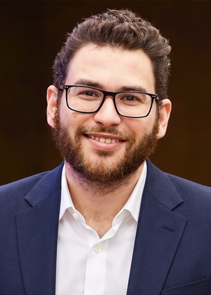
Perio-Implantologist
International Digital Dentistry Speaker
Digital Occlusion Consultant
Founder of DDS
Founder of Occlusa AI Platform
 h.sharshar@ddschoology.com
About Dr. Haitham Sharshar
Dr. Haitham Sharshar is an international digital dentistry speaker, educator, and digital occlusion consultant based in Cairo, Egypt. His work focuses on functional digital dentistry, CAD/CAM, implantology, jaw-motion analysis, and the integration of digital diagnostics into clinical treatment planning.
As Founder and Scientific Coordinator of Digital Dentistry Schoology (DDS), he has trained more than 3,270 dentists and dental technicians through university programs, international conferences, and hands-on courses. His teaching connects patient-specific diagnostic records with digital design and clinical workflows, helping clinicians and technicians understand how function and occlusion inform restorative planning.
Dr. Sharshar is a certified trainer for zebris JMA-Optic+ jaw-motion analysis and MyoWise dental EMG. His educational and consulting work brings together patient diagnostics, jaw-motion records, muscle-activity data, digital occlusion, and AI-supported workflows.
He is Founder and Clinical Director of Occlusa, an AI-supported platform for organizing clinical information and supporting clinician-led review and treatment planning. He also owns HS Dental Clinic in Cairo, where his clinical focus includes full-mouth digital rehabilitation and smile design.
His speaking and training topics include functional digital dentistry, digital occlusion, jaw-motion analysis, CAD/CAM workflows, digital implantology, and the practical integration of AI-supported tools into dental education and clinical practice.
Happy to collaborate on spreading digital dentistry.