Patient-Centered Aesthetic Treatment Planning in Cosmetic Prosthodontics: A Comprehensive Approach
A useful aesthetic treatment plan connects what the patient wants to change with what the teeth, supporting tissues and proposed restoration can reasonably accommodate. Photographs, scans and smile simulations can make that conversation more concrete. They do not, by themselves, establish a diagnosis, predict satisfaction or guarantee the final shade.
This DDS teaching guide follows that conversation from the first consultation to the laboratory prescription and review appointment. It distinguishes patient-reported outcomes from technical measurements and explains why a convincing digital preview still needs clinical assessment.
What matters to this patient?
Begin by asking which features the patient wants to change, which they want to preserve, and what would make treatment worthwhile. A request for a “better smile” may concern tooth colour, length, alignment, visible gingiva or a combination. Record the person’s own description before proposing a design. This is a practical consultation approach, not a validated formula for increasing treatment acceptance.
The American Dental Association’s consent guidance describes a discussion of the condition, proposed care, benefits, risks and alternatives, including no treatment. Give the patient an opportunity to ask questions and time to consider complex treatment. Local consent requirements still apply; a simulation or signed design approval does not replace the clinical discussion.
When a structured appearance assessment is useful, the Orofacial Esthetic Scale validation study by Larsson and colleagues provides a relevant example. The instrument assesses self-reported orofacial appearance in prosthodontic patients. Validation supports measuring the patient’s perspective; it does not show that administering the questionnaire causes a particular improvement in satisfaction. Use the appropriate validated language version and its scoring instructions rather than creating an apparent percentage benefit from unrelated scores.
How do preferences connect with clinical findings?
Use the consultation to identify questions that the examination must resolve. The following is an educational planning checklist, not a treatment prescription:
- Health and preservation: assess the teeth and periodontal tissues, existing restorations and the reason for any wear or damage before deciding whether an aesthetic intervention is appropriate.
- Function: record relevant concerns about chewing, speech, comfort and tooth contacts. An attractive preview cannot establish that a proposed restoration will function comfortably.
- Appearance: document tooth display, colour, form, alignment and gingival relationships in the context of the patient’s face and preferences. Measurements describe the starting point; they do not compel treatment to a fixed proportion.
- Practical choices: discuss the scope of treatment, maintenance, likely visits, cost and the uncertainty that matters to the individual. Consider a specialist opinion when the questions extend beyond the treating clinician’s competence.
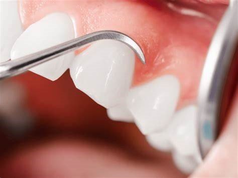
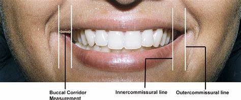
Keep the clinical findings distinct from the proposed design. For example, a request to lengthen the front teeth raises questions about restorative space, speech and function; it is not an automatic indication for veneers or crown lengthening. The plan should explain why any irreversible procedure is necessary and what reasonable alternatives were considered.
What can a preview demonstrate?
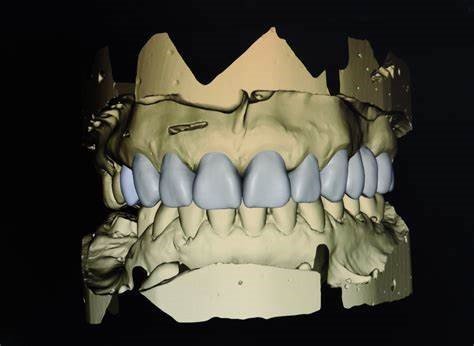
A two-dimensional smile simulation can help discuss visible tooth form and position. A three-dimensional design can add a spatial view of the proposed restorations. A diagnostic wax-up or intraoral mock-up offers another way to discuss the proposal, where clinically appropriate. These representations answer different questions and should be labelled clearly as proposed designs.
A published clinical report by Garcia and colleagues illustrates the distinction: the team used a direct mock-up when the digital simulation was insufficient for communicating the planned anterior rehabilitation. This is a traceable clinical example, not a comparative trial or evidence that every patient needs the same treatment. The images in this guide are separate illustrations, not photographs from that report.
Patient outcomes need equally careful interpretation. Luniyal and colleagues’ 2024 randomized trial enrolled 150 adults and reported higher immediate satisfaction in its digital smile-design group; professional assessments were made at three months. That finding concerns the tested planning approaches and time points. It does not establish long-term satisfaction, a benefit for every commercial system, or an effect caused by one questionnaire or preview feature alone.
During the preview discussion, ask what the patient likes, what feels unfamiliar and what still needs explanation. Record proposed changes and revisit them after an appropriate clinical try-in. A patient may reasonably decide against treatment. Acceptance is a decision, not proof that the plan is beneficial or that consent was adequately informed.
Does a colour scan establish the final restoration shade?
Shape capture, surface colour display and shade determination are different functions. A natural-looking scan is not automatically a calibrated shade measurement. Repeatability describes whether a device gives similar results when measurements are repeated; agreement with another method describes a different property. Neither measurement, on its own, demonstrates a better laboratory-made restoration or a happier patient.
In a clinical study by Ebeid and colleagues, 20 participants and 10 observers were used to compare a spectrophotometer and four intraoral scanners with visual shade selection. The scanners included TRIOS 3 and TRIOS 4, and CEREC Omnicam and Primescan. Most tested instruments performed similarly to the observers against the study’s visual reference; Omnicam performed less accurately than the other instruments. The reference was the most frequent visual shade choice, not an independent measurement of the final restoration’s colour.
The comparator also matters. Rutkūnas, Dirsė and Bilius compared TRIOS 3 with a spectrophotometer in 20 participants and found that shade readings did not exactly match, recommending additional shade-assessment methods. In a separate 31-participant study by Abu-Hossin and colleagues, TRIOS 3 and Omnicam showed only slight agreement with visual selection; the authors recommended visual confirmation. Different teeth, reference methods, shade systems and scanner generations limit direct comparisons between studies.
These studies support cautious use of scanner shade information as one part of assessment. They do not establish a universal improvement in laboratory shade communication over photography. As an educational application, the dentist and technician should agree on the tooth and region measured, shade system, capture method and supporting records. If visual and instrument findings disagree, resolve the discrepancy before relying on a single code. Examine the proposed restoration clinically rather than treating a screen colour as a final acceptance test.
Which tool performs the task you actually need?
Choose a tool by the clinical question and the information the laboratory can use. Check the exact hardware, software version, licensed modules, calibration requirements and local instructions. Product descriptions identify functions; they do not establish comparative clinical superiority.
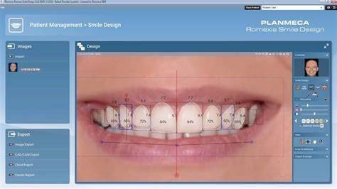
- Photo-based smile design: Planmeca Romexis Smile Design uses a two-dimensional face photograph and editable tooth silhouettes. The manufacturer’s description does not establish prediction of biological soft-tissue healing. Its exported design is a reference for subsequent work, not proof of final fit.
- Design and simulation software: Smile Designer Pro describes photo-based planning, simulation and CAD/CAM overlay functions. Confirm the current subscription, export options and compatibility for the intended workflow. Historical price tables and generic “one-time” prices should not determine a purchase.
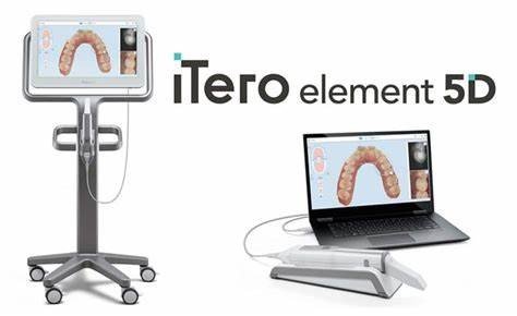
- Colour imaging and other imaging functions: the iTero Element 5D manufacturer describes three-dimensional, intraoral colour and near-infrared images. Those functions do not, by themselves, validate a quantitative restorative shade result. Evidence about interproximal caries detection is not evidence of shade-selection accuracy.
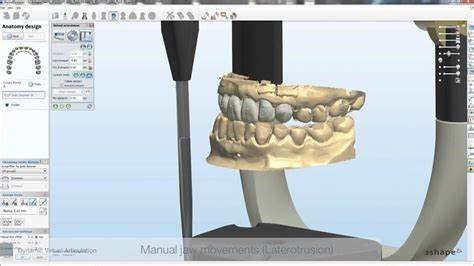
- Shade records sent with a scan: 3Shape’s Unite 23.1 guidance describes labelled shade screenshots and comments sent to the laboratory. It is version-specific workflow documentation, not a clinical accuracy trial or confirmation that every TRIOS configuration has identical features.
Before committing a case to a digital route, check a representative export with the receiving laboratory. Establish which records arrive, how the proposed design will be interpreted and which clinical information must accompany it. A colour model, a photograph and a numerical shade designation should not be treated as interchangeable records. Follow the relevant current device instructions rather than extrapolating settings from a different generation.
How should the plan be agreed and reviewed?
Bring the patient’s priorities, examination findings and proposed design back together. Explain what each option is intended to achieve and what it cannot promise. Discuss the implications of material and treatment choices for preservation, appearance, maintenance and future intervention. Use an understandable written summary and record unresolved questions rather than letting an attractive rendering settle them.
The laboratory prescription should identify the agreed design and any subsequent changes. As a teaching checklist, include the relevant photographs and scans, target appearance, the location and method of shade assessment, the restoration and material being prescribed, and the clinical questions still awaiting a try-in. Share patient records only through an appropriately authorized clinical route.
At review, assess appearance from the patient’s perspective alongside clinical findings. Where a validated patient-reported measure is used, follow its scoring method consistently. Record the timing of the assessment and any unresolved concerns. Immediate approval of a preview, satisfaction after treatment and longer-term oral-health-related quality of life are different outcomes; do not combine them into a single success percentage.
What should the dentist and laboratory take forward?
- For the dentist: the OES research supports structured measurement of perceived appearance, while consent guidance supports discussing options. Record the patient’s priorities and use appropriate assessment tools; neither step guarantees satisfaction.
- For the dentist: the published mock-up report shows how a physical trial can clarify a digital proposal. Use the representation that answers the unresolved clinical question and distinguish a proposed appearance from a promised result.
- For the laboratory: shade studies show that device readings depend on the tested method and comparator. Ask for the measurement context and supporting records, and flag conflicting shade information before proceeding.
- For both: version-specific documentation explains what software can transfer. Confirm the actual files and agreed design, then review the restoration clinically and collect the patient’s feedback separately from technical measurements.
Patient-centered planning is a continuing conversation grounded in diagnosis, understandable choices and honest uncertainty. Digital tools are most useful when the team can explain which question each tool answers and which decisions still require clinical judgment.
References and product documentation
- American Dental Association. Types of Consent. Professional guidance; local legal requirements vary.
- Larsson P, John MT, Nilner K, List T. Reliability and validity of the Orofacial Esthetic Scale in prosthodontic patients. International Journal of Prosthodontics. 2010;23(3):257–262. Validation study.
- Garcia PP, et al. Digital smile design and mock-up technique for esthetic treatment planning with porcelain laminate veneers. Journal of Conservative Dentistry. 2018;21(4):455–458. DOI: 10.4103/JCD.JCD_172_18. Clinical report.
- Luniyal C, et al. Assessment of Patient Satisfaction and Treatment Outcomes in Digital Smile Design vs. Conventional Smile Design: A Randomized Controlled Trial. Journal of Pharmacy and Bioallied Sciences. 2024;16(Suppl 1):S669–S671. DOI: 10.4103/jpbs.jpbs_928_23.
- Ebeid K, Sabet A, El Sergany O, Della Bona A. Accuracy and repeatability of different intraoral instruments on shade determination compared to visual shade selection. Journal of Esthetic and Restorative Dentistry. 2022;34(6):988–993. DOI: 10.1111/jerd.12884.
- Rutkūnas V, Dirsė J, Bilius V. Accuracy of an intraoral digital scanner in tooth color determination. Journal of Prosthetic Dentistry. 2020;123(2):322–329. DOI: 10.1016/j.prosdent.2018.12.020.
- Abu-Hossin S, et al. Comparison of digital and visual tooth shade selection. Clinical and Experimental Dental Research. 2023;9(2):368–374. DOI: 10.1002/cre2.721.
- 3Shape. Quick guide to the different TRIOS scanners; Unite 23.1: shade-measurement order-form guidance. Manufacturer identification and version-specific workflow information.
- Dentsply Sirona. Connect Software. Manufacturer workflow information for Primescan and Omnicam; regional availability varies.
- Planmeca. Romexis Smile Design. Manufacturer product description.
- Smile Designer Pro. Design and simulation functions. Manufacturer product description.
- Align Technology. iTero Element 5D imaging system. Manufacturer product description; model and market limits apply.