
Neuromuscular Dentistry & AI-Driven Functional Restorations
Surface electromyography, jaw-motion recording and digital occlusal analysis measure different aspects of function. Their outputs may inform a clinician’s assessment, but a measurement, an AI-generated suggestion and a demonstrated treatment benefit are different things.
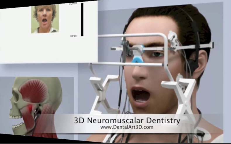
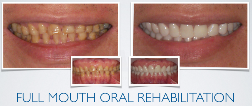
Clinical photographs and visualizations provide context; they do not establish a treatment effect or identify the cause of a patient’s symptoms.
What the tools measure
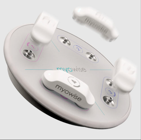
- Surface EMG: records muscle electrical activity. Myowise is an example of a wireless EMG system described by its manufacturer. Myowise product information.
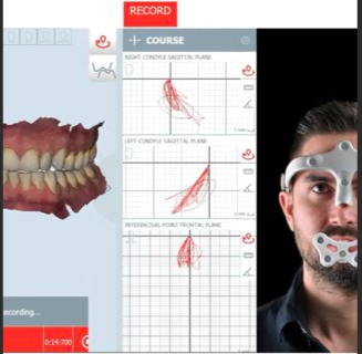
- Jaw-motion tracking: records mandibular movement. BioRESEARCH’s Bio JT-3D is an example of a jaw-tracking product. BioRESEARCH jaw tracker.
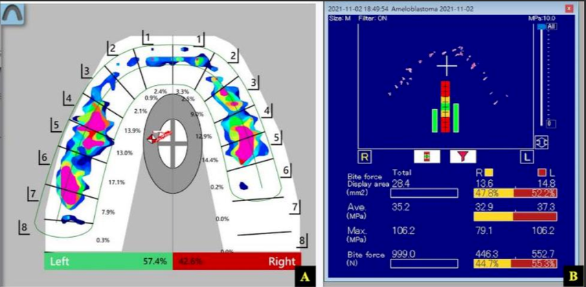
- Digital occlusal analysis: T-Scan records timing information during the bite sequence. The interpretation should follow the device documentation and the clinical examination. Tekscan: bite timing.
These manufacturer sources establish product identity and stated measurement capabilities. They are not independent proof that a device cures temporomandibular disorders or improves the survival of every restoration.
An important boundary for TMD
Jaw pain requires an appropriate diagnosis. NIDCR notes that evidence for many TMD treatments is limited and recommends conservative approaches. Irreversible changes to the teeth or bite should not be justified solely by an EMG trace, a jaw-motion recording or an occlusal display: evidence that occlusal treatments treat TMD is insufficient, and some can worsen the problem. NIDCR: temporomandibular disorders.
How to assess an AI claim
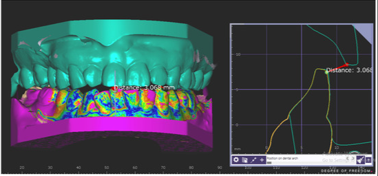
Before describing a workflow as AI-driven, identify the exact software, version and intended task. Ask whether the evidence concerns image processing, design assistance, contact analysis or patient outcomes. A colored CAD model does not establish that AI selected a clinically appropriate treatment.
For a claimed benefit, look for a traceable study that reports the patient group, comparison, endpoint and follow-up. A percentage improvement in “function” is not interpretable without knowing what was measured.
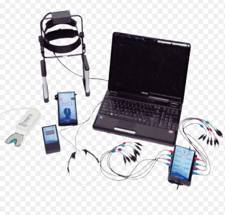 | 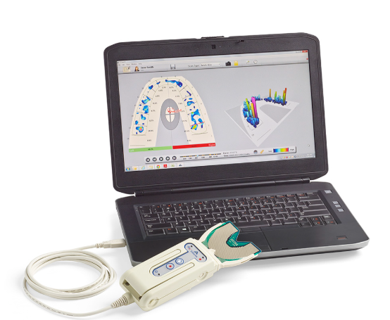 |
Equipment photographs are visual context, not evidence of clinical treatment benefit.
A practical evidence checklist
- Separate the clinical diagnosis from the instrument reading.
- Identify the exact device and software rather than a generic product label.
- Record how the findings affect a specific restorative decision.
- Check whether an apparent benefit comes from a controlled study, a case report or a manufacturer’s claim.
- Discuss uncertainty and treatment alternatives with the patient.
Sources
- National Institute of Dental and Craniofacial Research: TMD.
- BioRESEARCH: Bio JT-3D — manufacturer information.
- Myowise — manufacturer information.
- Tekscan: How to Analyze Bite Timing with T-Scan — manufacturer information.
Educational overview. Instrument readings and software suggestions do not replace clinical diagnosis.