
Multispectral Imaging in Oral Cancer Assessment: Evidence and Limits
Can an optical image decide whether an oral lesion needs biopsy? It cannot establish or exclude cancer. Clinical examination and tissue diagnosis remain central; a reassuring optical finding should not delay biopsy or specialist referral for a suspicious lesion. ADA guidance.
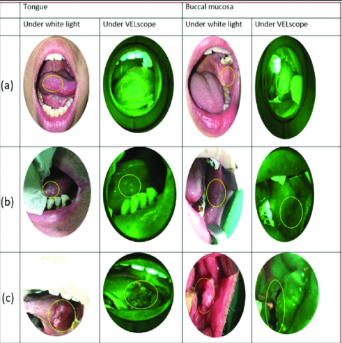
White-light and VELscope views show different appearances of oral tissues. Appearance alone does not establish a histopathological diagnosis. Read the diagnostic guidance.
What is multispectral imaging investigating?
Multispectral imaging (MSI) is being investigated for non-invasive visualization of oral tissue changes. Research systems analyze reflectance and/or fluorescence across selected wavelengths, but their performance depends on the device, algorithm, tissue site and study population. Preliminary imaging research should be distinguished from established clinical practice. Primary MSI study; spectral-imaging pilot study.
IARC estimated 389,846 new cases of lip and oral cavity cancer worldwide in 2022. The site and year matter when interpreting this estimate; prognosis varies with disease stage and patient characteristics. IARC GLOBOCAN 2022 fact sheet.
How do research systems collect tissue information?
- MSI records tissue responses in selected wavelength bands; the bands and imaging modes differ between systems.
- Research devices may combine reflectance and autofluorescence to investigate optical characteristics such as hemoglobin absorption and fluorescence changes.
- Image-analysis algorithms classify tissue in study datasets. Their accuracy and usefulness need validation for the intended device, population and setting.
These are research principles rather than universal device specifications. Bimodal MSI study; snapshot spectral-imaging study.
What have clinical studies actually shown?
A 2021 study of a bimodal multispectral system reported approximately 82% sensitivity and 96.6% specificity for distinguishing abnormal tissue (oral potentially malignant lesions plus squamous cell carcinoma) from patient-normal tissue across 89 sites. These are study-specific classification results, not a validated estimate of routine screening performance, fewer biopsies or improved survival. Read the primary study.
A separate early pilot described a snapshot spectral imaging system for oral reflectance and autofluorescence. Its exploratory findings support further investigation rather than routine clinical-outcome claims. Read the pilot study.
Is VELscope the same as a research MSI platform?
VELscope uses direct tissue autofluorescence to aid visualization of oral mucosal abnormalities. It is distinct from quantitative MSI research platforms. Manufacturer descriptions identify the imaging modality; they do not demonstrate improved survival, and an apparently reassuring fluorescence finding does not exclude cancer. ADA evidence summary.
What should change in clinical practice?
The ADA’s 2026 guideline conditionally recommends against light-based adjuncts for determining biopsy or referral needs. The recommendation for adults with mucosal abnormalities is conditional and based on very-low-certainty evidence. This recommendation concerns clinical use of light-based adjuncts; it does not declare every investigational MSI platform ineffective.
- Continue an appropriate visual and tactile examination and assess the history and observed mucosal abnormality.
- Arrange timely biopsy or specialist referral when a lesion is suspicious. Do not use an optical result to defer indicated care.
- Distinguish research classification accuracy from evidence that a device improves patient outcomes. Account for false-positive and false-negative results.
These clinical takeaways summarize current ADA guidance. NIDCR oral cancer information provides additional clinical and patient background.
What needs further research?
AI-assisted classification and handheld multispectral systems remain areas of investigation. As an educational interpretation of the evidence gaps, prospective validation should assess missed disease, downstream procedures and patient outcomes as well as diagnostic accuracy before routine benefits are claimed. Primary MSI research; pilot evidence.
The practical conclusion
MSI is an active research area. Promising image-classification results do not establish improved survival or justify replacing clinical examination and tissue diagnosis. The immediate priority for a suspicious lesion is timely biopsy or specialist referral, not reassurance from an optical image.
Sources and further reading
- ADA 2026 living guideline: light-based adjuncts and the need for biopsy.
- American Dental Association: Cancer (Head and Neck), Disease Detection section.
- Subhash and colleagues. Bimodal multispectral imaging system with cloud-based machine learning algorithm. Journal of Biomedical Optics. 2021;26(8):086003.
- Multimodal snapshot spectral imaging for oral cancer diagnostics: a pilot study.
- IARC GLOBOCAN 2022: Lip and oral cavity fact sheet.
- VELscope manufacturer technology description (device identity, not independent efficacy evidence).
- National Institute of Dental and Craniofacial Research: Oral Cancer.