
Impact of Parafunctional Habits on Prosthetic Success: Considering Occlusion and Functional Dynamics
How should bruxism change a prosthetic treatment plan? It warrants attention to the patient's activity, existing damage and restorative risks, but it does not identify one proven material, occlusal scheme or protective treatment. The useful distinction is between observed complications and evidence that an intervention prevents them.
What are you assessing: the behavior, damage or symptoms?
Sleep and awake bruxism are forms of masticatory muscle activity. The 2018 international consensus distinguishes that activity from its possible clinical consequences and discusses self-report and instrumental assessment. Describing the behavior, examining dental damage and assessing pain are related tasks, but they are not interchangeable findings.
Most of the research considered here concerns bruxism. It should not be generalized to every oral habit. For restorative planning, the question is which part of a study applies to this patient, this prosthesis and the outcome being discussed.
What does the evidence tell us about loading and complications?
Loading measurements do not predict an individual failure
In a small study of 10 participants, Nishigawa, Bando and Nakano (2001) measured nocturnal bite force over three nights and found that it could exceed maximum voluntary daytime bite force in some participants. These measurements describe nocturnal loading; they do not establish a ratio to ordinary chewing forces or predict prosthetic complication rates.
Long-term associations are not prevention trials
In a retrospective study from one specialist clinic, Chrcanovic, Kisch and Larsson (2020) evaluated 709 patients with implant-supported full-arch fixed prostheses over a mean follow-up of 10.7 years. Recorded bruxism was associated with more screw loosening and fracture, ceramic chipping or fracture, and prosthesis failure. These associations do not establish causation, prevention benefits or risks for all types of prosthesis.
This is a reason to discuss the findings in the context of full-arch implant prostheses. It is not a numerical risk estimate for every restoration, or proof that a splint, adjustment or material change will prevent a complication.
Laboratory material performance has a different meaning
In laboratory testing of specified three-unit fixed partial dentures, Heintze et al. (2018) found that fatigue resistance differed between the tested translucent zirconia and lithium disilicate materials and with zirconia connector size. This experiment measured mechanical fatigue performance; it did not measure clinical survival in patients with bruxism or establish a severity-based material prescription.
Before applying that comparison, identify whether the planned restoration resembles the tested material, three-unit design and connector configuration. A mechanical ranking from those specimens cannot be relabelled as a clinical survival ranking for people with bruxism.
How should those findings inform the plan?
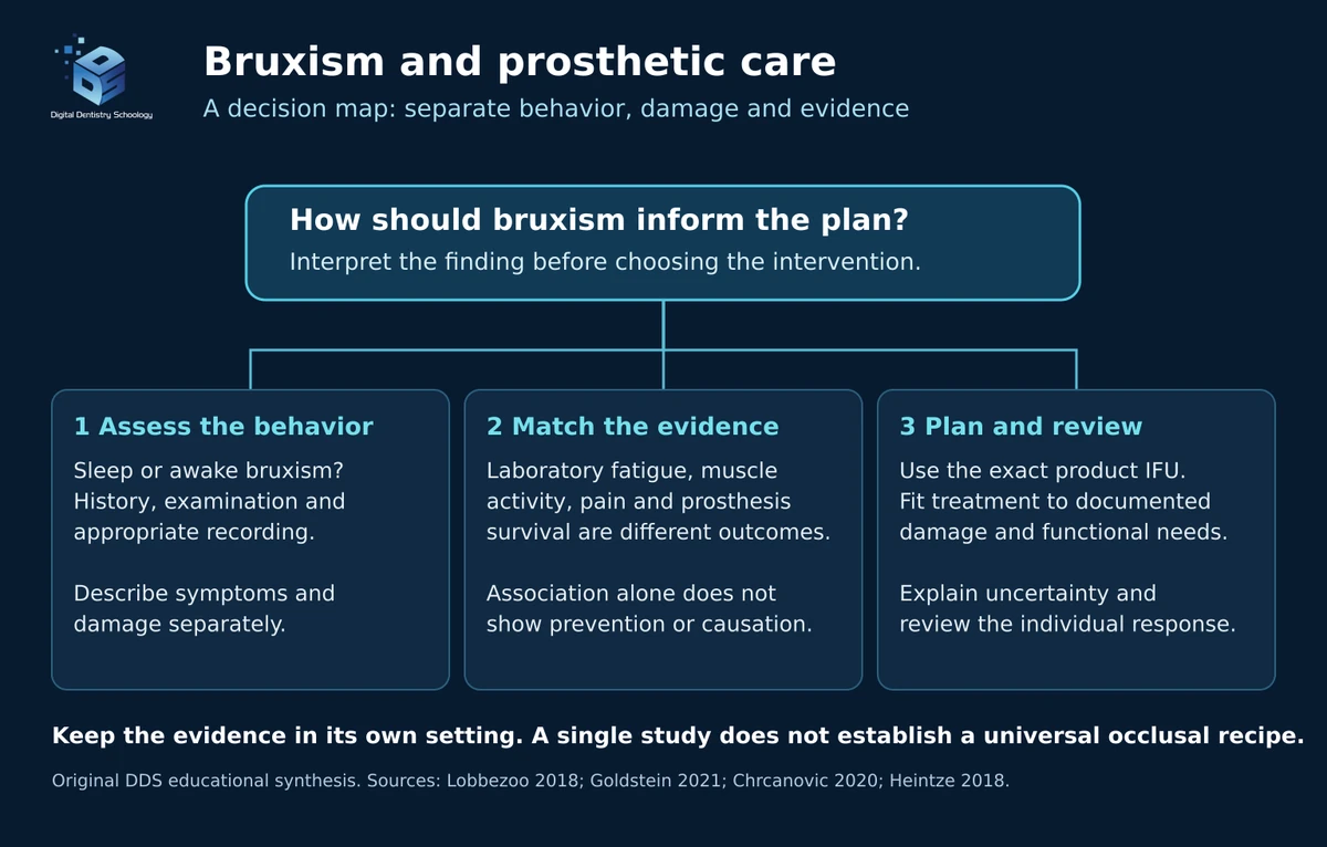
Three considerations belong together: what the patient is doing, what damage or symptoms are present, and what the cited evidence actually measured. These are questions to revisit during planning, not a validated sequence of treatments.
- Assess the behavior and consequences separately
Distinguish sleep from awake activity, and interpret history, examination and appropriate recordings in context. Describe damage and symptoms separately (Lobbezoo et al., 2018).
- Match the evidence to the intended outcome
Laboratory fatigue, muscle activity, pain and prosthesis survival answer different questions. The full-arch cohort reports associations; the three-unit experiment reports mechanical performance.
- Plan and review for the individual patient
Relate restorative choices to documented needs and exact product indications. Discuss the purpose of tooth protection, the limits of expected benefit and follow-up rather than promising a universal result.
Is there a proven occlusal scheme for bruxism?
The 2021 implant-restoration consensus found insufficient evidence to identify an occlusal scheme that minimizes complications. It does not establish a standard long-centric clearance, cusp angle, occlusal-table width or implant number for patients with bruxism.
- Cusp Angulation: A two-dimensional finite-element implant model found that cusp inclination and load position affected stress distribution. These findings do not establish a universal clinical cusp angle for patients with bruxism (Rungsiyakull et al., 2011).
- Group Function: An experiment in 10 healthy adults did not detect a difference in masseter electromyographic activity between devices using canine guidance or group function. It did not establish a preferred scheme for severe bruxism or test prosthetic longevity (Borromeo et al., 1995).
- Occlusal Vertical Dimension (OVD): The cited studies do not establish a bruxism-specific increase in vertical dimension or a predictable effect on muscle tension. Decisions about restorative space require an individual clinical indication.
These modeling and appliance-study findings do not establish a universal occlusal prescription or a percentage increase in restoration lifespan. Their populations, methods and measured outcomes need to be considered when applying them to restorative care.
Why does irreversible adjustment need a separate indication?
A 2023 randomized trial enrolled 17 healthy women with sleep bruxism; four were excluded from analysis. At follow-up averaging 14.4 months, it reported greater reductions in sleep muscle-activity episode frequency and duration after removal of occlusal interferences than after smoothing sharp margins without changing contacts.
This small study does not establish improved prosthetic survival or a general indication to reshape teeth for bruxism. Adjustment needed to correct a defined restorative problem is a separate clinical indication.
What can management reasonably aim to achieve?
Tooth protection, symptom management and restorative repair address different needs. NIDCR describes mouth guards as an option for separating and protecting teeth. That purpose should be distinguished from suppressing bruxism activity or extending prosthesis survival.
The table summarizes different kinds of evidence. It is not a head-to-head ranking, and the cited studies do not establish that one management package prevents peri-implant bone loss, eliminates headache or reduces treatment costs.
| Management Strategy | Effectiveness | Complexity | Patient Acceptance |
|---|---|---|---|
| Occlusal Splint Therapy | Used for tooth protection; a small four-week trial did not detect reduced sleep-bruxism activity at group level. | No common comparison | No common comparison |
| Biofeedback Training | One small, device-specific trial reported reduced sleep-bruxism bursts and facial-muscle pain during treatment. | No common comparison | No common comparison |
| Botulinum Toxin | Short-term effects in some trials; outcome-dependent | Clinical assessment and injections | Individual; discomfort or temporary weakness can occur |
| Occlusal Equilibration | Limited evidence; one small trial reported reduced sleep muscle activity | Irreversible if tooth structure is removed; indication-specific assessment | Not established by the cited trial |
| Medication (drug-specific) | Effects depend on the drug and outcome | Prescribing clinician assessment | Individual; review potential adverse effects |
| Cognitive Behavioral Therapy | Limited evidence; differs for sleep and awake bruxism | Structured behavioral work and follow-up | Individual |
| Material Modification | Laboratory fatigue performance varied by material and connector design; clinical survival in bruxism was not measured in that experiment. | No common comparison | No common comparison |
The splint, biofeedback and material-modification rows describe different outcomes: tooth protection, sleep-bruxism activity, facial-muscle pain and laboratory fatigue performance. They do not form a common ranking of clinical prosthesis survival. These studies report different outcomes and do not provide a shared comparison of complexity or patient acceptance.
Medication and behavioral evidence require outcome-specific interpretation
Medication and botulinum toxin treatment require individualized clinical assessment. Small placebo-controlled trials of amitriptyline did not show statistically significant reductions in sleep-bruxism muscle activity or associated pain (Mohamed et al., 1997; Raigrodski et al., 2001). Small trials of botulinum toxin type A have reported short-term reductions in selected measures of muscle activity, but effects on bruxism episodes and patient-reported symptoms have varied (Shim et al., 2020; Cruse et al., 2022). Temporary chewing weakness and injection discomfort have been reported.
A trial in patients with single-arch implant overdentures also recorded mechanical complications (Ali et al., 2021), but findings in that specific population do not establish protection for all prosthetic restorations. Treatment decisions should account for the clinical indication, potential adverse effects, and follow-up.
Evidence for behavioral approaches differs between sleep and awake bruxism. For sleep bruxism, a randomized trial found small improvements in both a multicomponent cognitive behavioral program and an occlusal-splint group, without significant between-group differences (Ommerborn et al., 2007). A systematic review found that evidence for CBT benefit in adult sleep bruxism remained limited (Minakuchi et al., 2022). For awake bruxism, a separate systematic review suggested possible benefits of behavioral approaches, while emphasizing limited and heterogeneous evidence (Graham et al., 2026).
Product indications are more useful than a catalogue ranking
A restorative ceramic and an occlusal-guard material serve different purposes. For example, the BruxZir Esthetic Milling Blanks instructions describe anterior and posterior restorations; they do not establish a guard design or suitability for severe bruxism. Use the current instructions for the exact product rather than transferring an indication or durability claim from another material.
Fit, use and response also need individual review. Adherence, changing activity patterns and the limits of assessment methods complicate follow-up; they should be discussed without implying that one contact scheme or appliance is optimal for everyone.
What is still being investigated?
Instrumented splints remain an area of research. Robin and colleagues reported home recordings over 10 consecutive nights using a wireless instrumented splint to characterize clenching and grinding events. This demonstrated monitoring capability in the studied setting; it did not show longer prosthesis survival. The device-specific biofeedback trial discussed above also does not establish that all wearables prevent damage or provide continuous protection.
What does one digital rehabilitation case show?
Stanley and colleagues reported a man with a chipped maxillary central-incisor veneer and tooth wear they attributed to bruxism. Initial records and a trial mock-up were made in 2015; definitive treatment resumed in 2017. This is a documented clinical and digital workflow, not an instrument-confirmed sleep-bruxism case or a controlled test of treatment efficacy.
For DDS readers, the value is seeing how clinical records, digital planning and delivery connect, while keeping that illustration separate from evidence that the treatment reduces bruxism or improves long-term survival.
The sequence below follows the report from initial assessment to treatment completion. The last clinical photographs and occlusal recording belong to the completion phase; they should not be mistaken for a six-month recall examination.
Choose a numbered step, swipe sideways, or use the Previous and Next links. Each step includes its clinical explanation and source figure.
Step 1 of 9
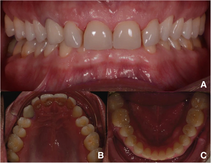
Baseline clinical photographs, 2015

Pretreatment frontal and upper/lower occlusal views. Source Figure 1; case credit and licence. The frontal and occlusal views document the dentition before rehabilitation. They support inspection of the reported wear and restorative problem; photographs alone do not quantify sleep or awake bruxism.
Step 2 of 9
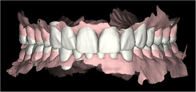
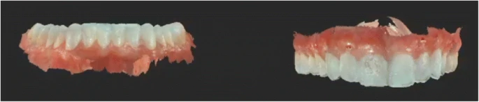
Initial digital record

Initial intraoral scan. Source Figure 3; case credit and licence. The initial intraoral scan records the upper and lower arches. In the reported workflow, those records were combined with facial and functional documentation for planning.
Step 3 of 9
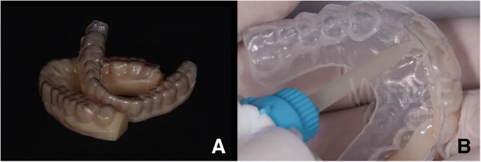
Models and trial mock-up

Model and matrix used to prepare the trial mock-up. Source Figure 5; case credit and licence. The authors used models and a matrix for a trial mock-up before definitive treatment. The reported trial illustrates how the proposed rehabilitation was explored; it does not establish a standard increase in vertical dimension.
Step 4 of 9
Records when treatment resumed, 2017

A new intraoral scan when treatment resumed after the two-year delay. Source Figure 8; case credit and licence. After an approximately two-year interruption, new intraoral records were obtained before treatment restarted. These are updated pretreatment records, not a two-year outcome.
Step 5 of 9
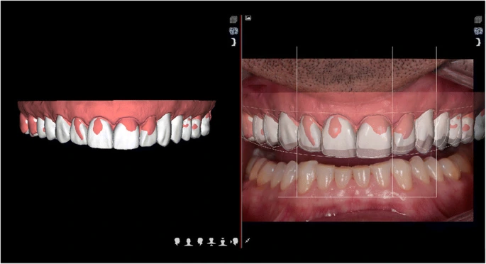
Digital planning

Digital Smile Design planning. Source Figure 11; case credit and licence. The digital planning view connects the clinical record to the proposed restoration design. It documents this team's design stage; it does not by itself establish improved clinical survival.
Step 6 of 9
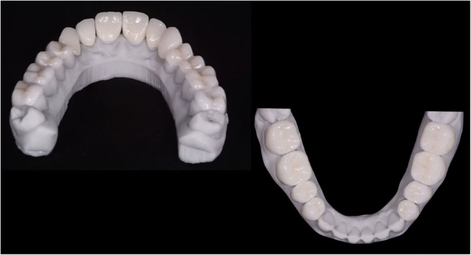
Fabricated restorations
Definitive restorations produced by the reported digital design and milling workflow. Source Figure 12; case credit and licence. The restorations are shown on laboratory models. The report describes digital design and milling. The material inconsistency discussed below prevents using this case as a material-selection or bonding protocol.
Step 7 of 9
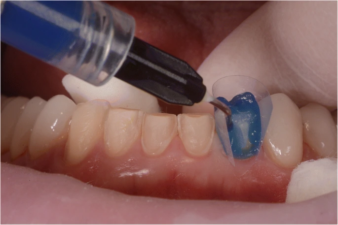
Adhesive delivery

A stage of adhesive restoration delivery from the published case. Source Figure 13; case credit and licence. The report describes a trial insertion to assess marginal fit and appearance before adhesive delivery. This close-up illustrates the delivery phase. Product-specific surface treatment and bonding values should come from the correct current instructions.
Step 8 of 9
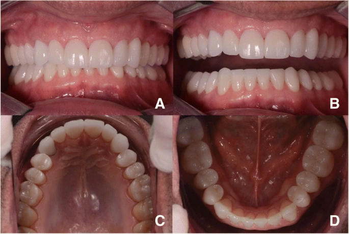
Post-treatment clinical views

Post-treatment photographs; the publication does not identify these as six-month recall images. Source Figure 15; case credit and licence. The frontal and occlusal views document the restored dentition after treatment. They are an observed post-treatment result, not evidence of long-term durability.
Step 9 of 9
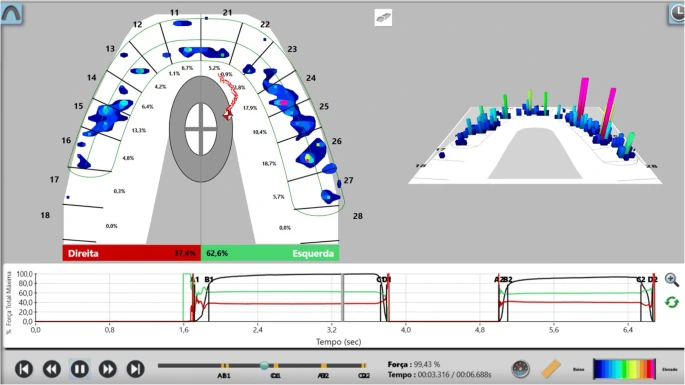
Occlusal recording at treatment completion

T-Scan recording used during the reported treatment. This does not measure bruxism diagnosis or cure. Source Figure 17; case credit and licence. The report describes occlusal adjustment and a T-Scan recording during completion, followed by an acrylic protective appliance. Relative contact-force and timing information does not establish a bruxism diagnosis, reduction in activity or cure.
What was reported at six months?
The authors reported that the restorations remained stable without fracture and that the patient reported no headaches. The six-month observation is described in text; no image is identified as a six-month recall photograph. These findings in one patient do not establish a causal effect on headache, temporomandibular disorders or bruxism activity, or survival beyond that observation. Stanley et al., case presentation.
Clinical Recommendations
For the dentist
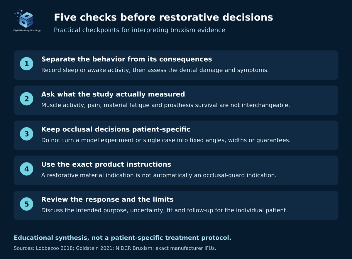
Separate the indication from the behavior. Describe sleep or awake activity, damage and symptoms before deciding what needs treatment. Bruxism assessment does not itself prescribe a restoration or a particular occlusal adjustment (assessment consensus; implant occlusal consensus).
Explain the outcome you are trying to influence. Tooth protection, pain relief and prosthesis survival are different goals. Use the table's source-specific findings to explain the limits of a proposed approach, rather than presenting a reduction in muscle activity as proof of longer restoration survival.
Keep the published case in proportion. Stanley's report illustrates records, planning and delivery for one patient. Its six-month observation cannot demonstrate that changing vertical dimension cures bruxism or establishes a generally successful restorative protocol.
For the dentist and laboratory
Identify the exact material and tested design. Compare a laboratory result with the planned prosthesis and connector configuration before applying it. The Heintze experiment supports interpretation of its tested specimens, not a severity-based clinical prescription.
Resolve material identity before choosing the processing instructions. The VITABLOCS classification mismatch shows why a paper's product label must be checked against the manufacturer's exact product information. A restorative-material indication also cannot be transferred to an occlusal guard.
A useful plan makes its uncertainties explicit and reviews the individual response. None of the cited evidence supplies a universal cusp angle, guard-material ranking or guaranteed increase in prosthesis lifespan.
References
- Rungsiyakull C, Rungsiyakull P, Li Q, Li W, Swain M. Effects of occlusal inclination and loading on mandibular bone remodeling: a finite element study. Int J Oral Maxillofac Implants. 2011;26(3):527-537.
- Goldstein G, Goodacre C, Taylor T. Occlusal Schemes for Implant Restorations: Best Evidence Consensus Statement. J Prosthodont. 2021;30(S1):84-90. doi: 10.1111/jopr.13319.
- Borromeo GL, Suvinen TI, Reade PC. A comparison of the effects of group function and canine guidance interocclusal device on masseter muscle electromyographic activity in normal subjects. J Prosthet Dent. 1995;74(2):174-180. doi: 10.1016/S0022-3913(05)80183-0.
- de Abreu T, Bahia C, Foscaldo T, et al. Effect of occlusal equilibration on masticatory muscle activity in females with sleep bruxism: a double-blind randomised controlled trial. J Sleep Res. 2023;32(4):e13879. doi: 10.1111/jsr.13879.
- Prismatik Dentalcraft, Inc. BruxZir Esthetic Milling Blanks: Instructions for Use. IFU-012562_1 (2021-01-20), page 1, sections 1-2. Accessed October 8, 2026.
- Lobbezoo F, Ahlberg J, Raphael KG, et al. International consensus on the assessment of bruxism: Report of a work in progress. J Oral Rehabil. 2018;45(11):837-844. doi: 10.1111/joor.12663.
- Nishigawa K, Bando E, Nakano M. Quantitative study of bite force during sleep associated bruxism. J Oral Rehabil. 2001;28(5):485-491. doi:10.1046/j.1365-2842.2001.00692.x.
- Chrcanovic BR, Kisch J, Larsson C. Retrospective evaluation of implant-supported full-arch fixed dental prostheses after a mean follow-up of 10 years. Clin Oral Implants Res. 2020;31(7):634-645. doi: 10.1111/clr.13600.
- Heintze SD, Monreal D, Reinhardt M, et al. Fatigue resistance of all-ceramic fixed partial dentures - Fatigue tests and finite element analysis. Dent Mater. 2018;34(3):494-507. doi: 10.1016/j.dental.2017.12.005.
- van der Zaag J, Lobbezoo F, Wicks DJ, et al. Controlled Assessment of the Efficacy of Occlusal Stabilization Splints on Sleep Bruxism. J Orofac Pain. 2005;19(2):151-158. doi: 10.11607/jofph.19151.
- Bergmann A, Edelhoff D, Schubert O, Erdelt KJ, Pho Duc JM. Effect of treatment with a full-occlusion biofeedback splint on sleep bruxism and TMD pain: a randomized controlled clinical trial. Clin Oral Investig. 2020;24(11):4005-4018. doi: 10.1007/s00784-020-03270-z.
- National Institute of Dental and Craniofacial Research. Bruxism. Treatment: Mouth Guards. Last reviewed March 2025; accessed October 8, 2026.
- Mohamed SE, Christensen LV, Penchas J. A randomized double-blind clinical trial of the effect of amitriptyline on nocturnal masseteric motor activity (sleep bruxism). Cranio. 1997;15(4):326-332. doi: 10.1080/08869634.1997.11746027.
- Raigrodski AJ, Mohamed SE, Gardiner DM. The effect of amitriptyline on pain intensity and perception of stress in bruxers. J Prosthodont. 2001;10(2):73-77. doi: 10.1111/j.1532-849x.2001.00073.x.
- Shim YJ, Lee HJ, Park KJ, et al. Botulinum Toxin Therapy for Managing Sleep Bruxism: A Randomized and Placebo-Controlled Trial. Toxins (Basel). 2020;12(3):168. doi: 10.3390/toxins12030168.
- Cruse B, Dharmadasa T, White E, et al. Efficacy of botulinum toxin type a in the targeted treatment of sleep bruxism: a double-blind, randomised, placebo-controlled, cross-over study. BMJ Neurol Open. 2022;4(2):e000328. doi: 10.1136/bmjno-2022-000328.
- Ali SM, Alqutaibi AY, Aboalrejal A, Elawady DM. Botulinum toxin and occlusal splints for the management of sleep bruxism in individuals with implant overdentures: A randomized controlled trial. Saudi Dent J. 2021;33(8):1004-1011. doi: 10.1016/j.sdentj.2021.07.001.
- Ommerborn MA, Schneider C, Giraki M, et al. Effects of an occlusal splint compared with cognitive-behavioral treatment on sleep bruxism activity. Eur J Oral Sci. 2007;115(1):7-14. doi: 10.1111/j.1600-0722.2007.00417.x.
- Minakuchi H, Fujisawa M, Abe Y, et al. Managements of sleep bruxism in adult: A systematic review. Jpn Dent Sci Rev. 2022;58:124-136. doi: 10.1016/j.jdsr.2022.02.004.
- Robin O, Claude A, Gehin C, Massot B, McAdams E. Recording of bruxism events in sleeping humans at home with a smart instrumented splint. Cranio. 2022;40(1):14-22. Published online January 8, 2020. doi: 10.1080/08869634.2019.1708608.
- Graham DA, Lövgren A, Häggman-Henrikson B, et al. Management of awake bruxism: a systematic review. BMC Oral Health. 2026;26:559. doi: 10.1186/s12903-026-07856-z.
- Stanley M, Paz AG, Miguel I, Coachman C. Fully digital workflow, integrating dental scan, smile design and CAD-CAM: case report. BMC Oral Health. 2018;18:134. doi: 10.1186/s12903-018-0597-0.
- VITA Zahnfabrik. VITABLOCS TriLuxe forte: product information. Manufacturer identification of the named feldspar ceramic. Accessed October 9, 2026.
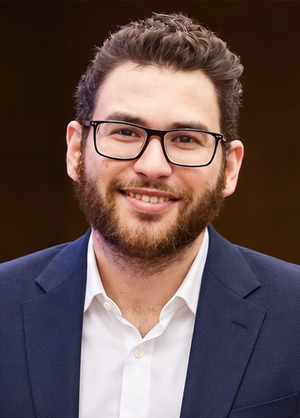


About Dr. Haitham Sharshar
Dr. Haitham Sharshar is an international digital dentistry speaker, educator, and digital occlusion consultant based in Cairo, Egypt. His work focuses on functional digital dentistry, CAD/CAM, implantology, jaw-motion analysis, and the integration of digital diagnostics into clinical treatment planning.
As Founder and Scientific Coordinator of Digital Dentistry Schoology (DDS), he has trained more than 3,270 dentists and dental technicians through university programs, international conferences, and hands-on courses. His teaching connects patient-specific diagnostic records with digital design and clinical workflows, helping clinicians and technicians understand how function and occlusion inform restorative planning.
Dr. Sharshar is a certified trainer for zebris JMA-Optic+ jaw-motion analysis and MyoWise dental EMG. His educational and consulting work brings together patient diagnostics, jaw-motion records, muscle-activity data, digital occlusion, and AI-supported workflows.
He is Founder and Clinical Director of Occlusa, an AI-supported platform for organizing clinical information and supporting clinician-led review and treatment planning. He also owns HS Dental Clinic in Cairo, where his clinical focus includes full-mouth digital rehabilitation and smile design.
His speaking and training topics include functional digital dentistry, digital occlusion, jaw-motion analysis, CAD/CAM workflows, digital implantology, and the practical integration of AI-supported tools into dental education and clinical practice.
Happy to collaborate on spreading digital dentistry.
h.sharshar@ddschoology.com
WHERE DIGITAL MEETS CLINICAL