
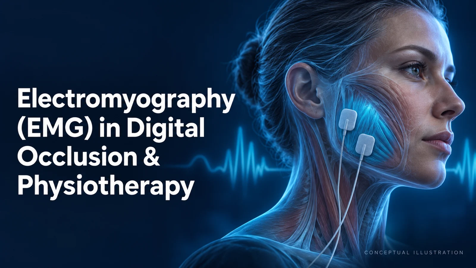
Electromyography (EMG) in Digital Occlusion & Physiotherapy
How should a clinician use an EMG trace when a patient reports jaw fatigue, clenching or discomfort during restorative treatment? Surface electromyography (sEMG) can describe muscle activation during a recorded task and support selected biofeedback or research applications. It does not, by itself, identify the cause of pain, measure force at each tooth or select a therapeutic bite position. Its value depends on the clinical question, the recording method and whether the result changes a justified management decision.
Learning objectives
After reading this article, you should be able to:
- Distinguish a surface EMG muscle-activation recording from a diagnosis, a tooth-force measurement and a treatment outcome.
- Identify recording conditions that must be documented before comparing EMG traces.
- Explain where EMG assessment and biofeedback may fit within a broader clinical evaluation of bruxism or persistent jaw pain.
- Recognize the limits of AI-assisted movement classification and choose patient-centred outcomes for follow-up.
The useful distinction is between recording a signal, interpreting a condition and improving a patient’s health. Those are separate steps. A more symmetrical trace is not automatically a healthier joint, a better restoration or evidence that treatment has succeeded. The measurement principles and clinical framework below explain how to keep those steps connected without confusing them.
What surface EMG measures
Surface electrodes detect electrical activity associated with muscle activation. Dental recordings commonly examine the superficial masseter and anterior temporalis muscles. Surface recordings and invasive needle or fine-wire recordings are different methods: an electrode on the cheek should not be described as a selective recording of every deep jaw muscle. Electrode location, skin contact, movement artefact and activity from adjacent muscles influence the signal. See Konrad’s technical introduction to EMG.
Muscle activation and force can be related under controlled conditions, but they are not interchangeable measurements. In a study of 84 people, Gonzalez and colleagues recorded sEMG alongside a separate calibrated bite-force transducer during defined static bites. The resulting relationships supported mechanical modelling for the tested biting conditions. They did not provide a chairside map of absolute force on every tooth or validate an EMG-selected full-mouth reconstruction.
A larger signal also does not establish muscle fatigue. Experimental fatigue analysis can examine changes in amplitude and frequency during specified tasks; the conditions matter. An isolated high reading during chewing or clenching is insufficient to label a muscle fatigued or to prescribe a bite change. This distinction follows the task-dependent interpretation described in Konrad’s fatigue-analysis chapter.
Start with the complaint, then choose the record
For jaw pain or restricted function, take a history, examine the patient and consider competing causes before interpreting a dashboard. The Diagnostic Criteria for Temporomandibular Disorders (DC/TMD) combine a clinical assessment of common pain-related TMD with assessment of pain-related disability, jaw function and psychosocial factors. This framework should not be replaced by an EMG asymmetry score. Some joint diagnoses require further investigation beyond the clinical screening criteria.
Occlusion describes how teeth contact. It is not an established explanation for every painful jaw. NIDCR’s TMD guidance states that current evidence does not support a strong link between malocclusion and TMD. It also warns that changing teeth or the bite to treat TMD has insufficient evidence of benefit and can worsen symptoms. That warning concerns treatment of TMD; it does not remove an independent indication to repair a damaged tooth or a defective restoration.
In full-mouth rehabilitation, therefore, document the restorative problem and assess contacts, jaw function, symptoms and the planned restorations with the appropriate records. If sEMG is added, state the narrower question it addresses, such as whether a recorded activation pattern is reproducible. Do not grind teeth, change vertical dimension or redesign a restoration merely to make the two sides of a graph match. This is a clinical reasoning safeguard, not a validated EMG treatment protocol.
Make before-and-after recordings comparable
A useful repeat recording needs a reproducible task and a record of how it was collected. Sforza and colleagues studied 40 healthy young adults using specified clenching and shoulder-elevation tasks. Reproducibility varied with the measurement index. Those results support attention to protocol and repeatability; they do not establish universal diagnostic thresholds for painful TMD or a treatment target for every patient.
- Define the question: record whether the aim is task comparison, monitoring or learning a relaxation response.
- Record the setup: include muscles, electrode sites, device and processing settings, posture, task, reference or normalization method and symptoms during testing.
- Check an unexpected trace: inspect electrode contact and artefact, then repeat the same task before interpreting a change.
- Compare like with like: a different task, recording setup or reference contraction can change the meaning of the numbers.
These are practical recording checks drawn from EMG acquisition principles, not a universal device prescription. In a painful or injured patient, do not force a maximal clench simply to obtain a reference value; choose an appropriate assessment with the treating clinician.
Bruxism assessment: activity and consequences
Bruxism includes jaw-muscle activity during sleep and wakefulness, and extends beyond audible tooth grinding. The 2025 international consensus update distinguishes self-reported, clinically assessed and device-assessed information rather than treating them as a simple diagnostic hierarchy. Specify which type of evidence is available. A patient’s report, tooth-wear finding and overnight muscle recording answer different questions.
EMG can contribute to device-based assessment, but an activity count alone does not show that the person has pain, ongoing tooth damage or a need for intervention. Sleep-related activity can fluctuate between recordings. Interpret it alongside the patient’s symptoms, examination and relevant sleep or medical history. The same consensus advises managing adverse consequences when indicated, and investigating an underlying disorder when bruxism is a sign of one. It does not justify calling all bruxism a disease caused by a misaligned bite.
Where physiotherapy and biofeedback fit
Biofeedback gives a person information about a physiological signal while they learn a response. NIDCR describes therapist-assisted use of jaw-muscle sensing to help a patient notice tightening and practise relaxing. The sensor supplies feedback; the treatment includes learning and clinical supervision. A passive recording or a wearable alert is not necessarily the same intervention. See the NIDCR behavioral-treatment explanation.
For adults with moderate chronic TMD pain lasting at least three months, the 2023 BMJ clinical practice guideline favours interventions including supervised jaw exercise and stretching, therapist-assisted mobilisation, usual care and cognitive behavioural therapy (CBT) with or without biofeedback or relaxation. It conditionally recommends against biofeedback used on its own. The guideline does not address acute TMD pain, and its recommendations do not validate every app, sensor or exercise programme.
A relevant randomized trial by Shedden Mora and colleagues compared eight weekly sessions of biofeedback-based CBT with eight weeks of occlusal-splint treatment in 58 people with chronic TMD. Both groups improved in pain and disability; pain-coping improvement favoured the combined psychological treatment. No significant change in nocturnal masseter activity was observed. The study therefore illustrates why symptom improvement and EMG change should be assessed separately. It does not establish the effect of biofeedback alone.
What AI-assisted EMG has actually demonstrated
Machine learning can classify patterns in recorded signals, but its performance is specific to the data, movements and people tested. In a 2026 experiment by Minakuchi and colleagues, 12 healthy participants performed simulated bruxism and non-bruxism movements while awake. The models were trained on 11 participants and tested on the remaining participant in turn. Masseter recordings helped distinguish simulated tooth-contact movements, but performance for simulated movements without tooth contact was poor.
This was a small laboratory classification study, not a trial of overnight diagnosis, pain treatment or autonomous restoration adjustment. The authors identified validation with actual sleep EMG and polysomnography data as future work. Its findings cannot support a general claim that AI sensors prevent tooth wear, cure TMD or automatically determine a better bite.
Before using an automated label in care, ask what it was tested against, whether the study population and recording conditions match the patient, which events it misses, and who reviews uncertain results. A useful label may prompt a closer look at a recording. A recommendation to alter treatment requires separate clinical evidence and an accountable clinician.
Judge follow-up by the patient as well as the trace
Agree on the outcome before collecting more data: less pain during meals, improved jaw function, better coping or a separately defined restorative goal. Record symptoms, function, adverse effects and relevant examination findings alongside any EMG measures. If the trace changes but the patient does not improve, reassess the diagnosis and management rather than chasing a numerical target. DC/TMD’s function and disability assessments and the biofeedback-CBT trial provide the rationale for that distinction.
EMG is most useful when it answers a defined measurement question or supports a specific learning task. Digital records can make observations easier to review; the clinical assessment determines what those observations mean and whether a proposed intervention is justified.
A map for reading an EMG record
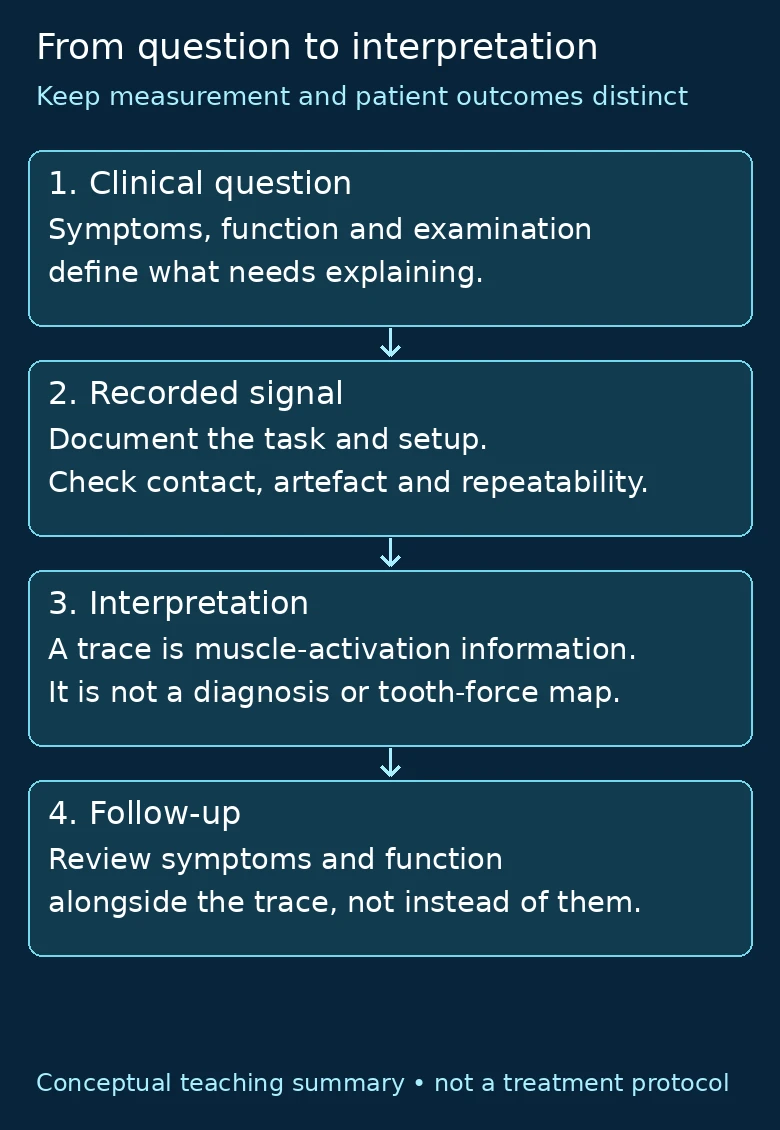
- Clinical question: symptoms, function and examination define what needs explaining.
- Recorded signal: document the task and setup; check contact, artefact and repeatability.
- Interpretation: a trace provides muscle-activation information, not a diagnosis or a map of tooth force.
- Follow-up: review symptoms and function alongside the trace, rather than replacing them with it.
The links between these stages follow the article’s discussion of EMG acquisition principles, DC/TMD assessment, activation and separately measured bite force, and the biofeedback-CBT trial’s distinction between symptoms and EMG change.
Three interpretation checks
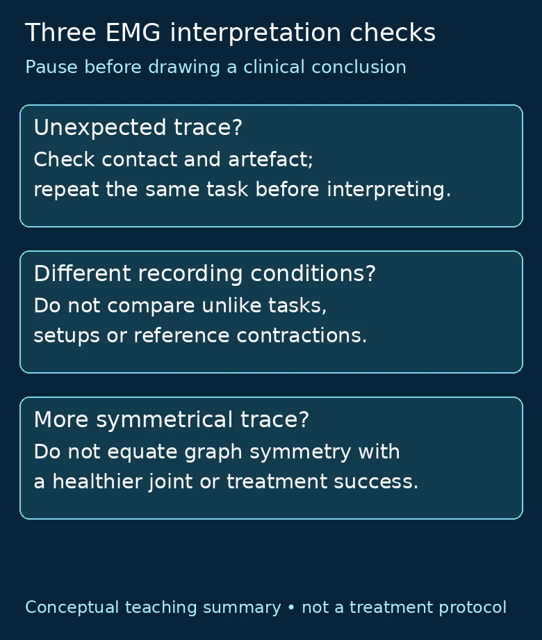
- Unexpected trace? Check electrode contact and artefact, then repeat the same task before interpreting the change.
- Different recording conditions? Do not treat unlike tasks, setups or reference contractions as equivalent comparisons.
- More symmetrical trace? Do not equate graph symmetry with a healthier joint or treatment success.
These reminders condense the existing recording-comparability and clinical-interpretation sections, drawing on Konrad’s technical guidance, Sforza and colleagues’ protocol-specific reproducibility study, and the article’s NIDCR-supported caution about bite changes for TMD.
References
View the cited technical guidance and clinical research
- Konrad P. The ABC of EMG: A Practical Introduction to Kinesiological Electromyography. Version 1.4, March 2006. Noraxon.
- Gonzalez Y, et al. Reliability of electromyographic activity vs. bite-force from human masticatory muscles. European Journal of Oral Sciences. 2011;119:219–224.
- Schiffman E, et al. Diagnostic Criteria for Temporomandibular Disorders (DC/TMD) for Clinical and Research Applications. Journal of Oral & Facial Pain and Headache. 2014;28:6–27.
- National Institute of Dental and Craniofacial Research. TMD (Temporomandibular Disorders). Reviewed September 2026.
- Sforza C, et al. EMG analysis of trapezius and masticatory muscles: experimental protocol and data reproducibility. Journal of Oral Rehabilitation. 2011;38:648–654.
- Verhoeff MC, et al. Updating the Bruxism Definitions: Report of an International Consensus Meeting. Journal of Oral Rehabilitation. 2025;52:1335–1342.
- Busse JW, et al. Management of chronic pain associated with temporomandibular disorders: a clinical practice guideline. BMJ. 2023;383:e076227.
- Shedden Mora MC, et al. Biofeedback-based cognitive-behavioral treatment compared with occlusal splint for temporomandibular disorder: a randomized controlled trial. Clinical Journal of Pain. 2013;29:1057–1065.
- Minakuchi H, et al. Experimental Analysis of Automatic Discrimination Performance Between Simulated Bruxism and Non-Bruxism Under Conscious Conditions Using Electromyography and Machine Learning. International Journal of Dentistry. 2026;2026:7874254.
About Dr. Haitham Sharshar



Dr. Haitham Sharshar is an international digital dentistry speaker, educator, and digital occlusion consultant based in Cairo, Egypt. His work focuses on functional digital dentistry, CAD/CAM, implantology, jaw-motion analysis, and the integration of digital diagnostics into clinical treatment planning.
As Founder and Scientific Coordinator of Digital Dentistry Schoology (DDS), he has trained more than 3,270 dentists and dental technicians through university programs, international conferences, and hands-on courses. His teaching connects patient-specific diagnostic records with digital design and clinical workflows, helping clinicians and technicians understand how function and occlusion inform restorative planning.
Dr. Sharshar is a certified trainer for zebris JMA-Optic+ jaw-motion analysis and MyoWise dental EMG. His educational and consulting work brings together patient diagnostics, jaw-motion records, muscle-activity data, digital occlusion, and AI-supported workflows.
He is Founder and Clinical Director of Occlusa, an AI-supported platform for organizing clinical information and supporting clinician-led review and treatment planning. He also owns HS Dental Clinic in Cairo, where his clinical focus includes full-mouth digital rehabilitation and smile design.
His speaking and training topics include functional digital dentistry, digital occlusion, jaw-motion analysis, CAD/CAM workflows, digital implantology, and the practical integration of AI-supported tools into dental education and clinical practice.
WHERE DIGITAL MEETS CLINICAL
Happy to collaborate on spreading digital dentistry.
h.sharshar@ddschoology.com