
Color Matching Strategies in Anterior Restorations: The Art and Science of Aesthetic and Cosmetic Prosthodontics
Why can an anterior restoration carry the correct shade label yet still look different from its neighbour? A shade code describes only part of the problem. Lighting, tooth hydration, the region measured, the underlying tooth and the restorative material all influence the result. A useful strategy combines a reproducible shade record with visual assessment and checks the restoration itself before definitive acceptance.
This guide connects colour science, measurement evidence and laboratory communication. Its practical checkpoints are educational guidance for clinician-led care; a device reading or a favourable study result cannot guarantee an individual patient’s aesthetic outcome.
What needs to match?
Colour perception involves the light source, the object and the observer. Hue describes a colour family, value its lightness, and chroma its intensity. CIELAB describes lightness with L*, the red–green component with a*, and the yellow–blue component with b*. These coordinates are useful records, but they do not describe the whole appearance of an anterior tooth.


Translucency, surface texture and gloss also matter. Enamel and dentine form a layered optical structure, so a uniform shade tab cannot reproduce every cervical, middle and incisal feature. Opalescence and fluorescence concern how light interacts with the tissues or material; neither should be inferred from an ordinary photograph alone. Sikri’s colour-science review provides background terminology, rather than comparative evidence that one product produces a better restoration.
Build a reproducible shade record
Begin with the conditions under which the shade is recorded. In a prospective study of 25 participants with intact maxillary incisors, Alamé and Mehanna Zogheib found that rubber-dam dehydration changed measured colour and increased lightness during the observation period. The finding supports recording hydration status and avoiding a dehydrated tooth as an unquestioned colour target. It does not establish one rehydration interval for every clinical situation.
- Record the starting conditions. Note the reference tooth, lighting, hydration and any existing restorations or discolouration that could affect the target.
- Keep visual and instrumental records connected. State the shade-guide system and exact device model. Follow that device’s current calibration, positioning and infection-control instructions.
- Describe regional differences. A measurement from the middle third is a local reading. Add a cervical-to-incisal description when the restoration must reproduce a gradient; do not assume that one reading represents the whole tooth.
- Use photographs as communication aids. Record the lighting and calibration method when interpreting colour from images. An ordinary phone photograph, a smile preview and a calibrated colour-measurement system are different records.
- Repeat a doubtful acquisition. Recheck positioning, calibration, tooth condition and the selected shade scale before changing the restorative prescription because of a conflicting result.
These checkpoints are a practical synthesis. They make the record easier to interpret; they are not a separately validated protocol or a promise of fewer remakes.
What do device comparisons show?
Repeatability is not the same as accuracy or patient satisfaction. Repeatedly choosing the same shade shows consistency. Agreement with another method, difference from a reference colour, the appearance of a completed crown and a patient’s acceptance are separate outcomes.
In Liberato et al.’s clinical comparison, three clinicians and two instrumental methods assessed the middle third of a maxillary central incisor in 28 participants under controlled lighting. The tested TRIOS scanner and VITA Easyshade Advance 4.0 showed greater reliability than the tested visual methods. Results depended on the shade scale. This supports those methods under those conditions, rather than a universal percentage improvement or a ranking of every current scanner and spectrophotometer.
The genuine Dozić et al. device study was published in 2007. It compared five instruments on shade tabs and on central incisors in 25 dental students. Device performance differed between laboratory and clinical conditions, and clinical accuracy did not differ statistically among ShadeScan, Ikam and Easyshade. The instruments and generations tested cannot establish a present-day league table for Easyshade V, SpectroShade Micro, Rayplicker or smartphone systems.
There is also evidence about fabricated restorations. Paul et al. compared visual and spectrophotometric workflows for porcelain-fused-to-metal incisor crowns in 10 patients and reported better colour matching with the spectrophotometric workflow. This small, material-specific clinical comparison is not a long-term survival study and should not be transferred directly to every composite or all-ceramic restoration.
For decision-making, ask what a study measured, against which reference, on which tooth or specimen, with which device generation and over what period. Do not combine unlike colour formulas, reliability coefficients and clinical preference scores into one “accuracy” percentage.
Choose a measurement tool for the task
The following examples explain functions, not comparative superiority. Check the current instructions, service support, software compatibility and local quotation for the exact configuration being considered.
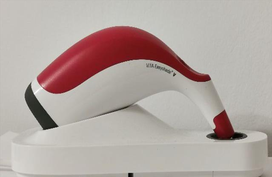
VITA Easyshade V
VITA identifies Easyshade V as a spectrophotometer for natural teeth and ceramic restorations, with shade-system outputs and L*a*b*/L*C*h values. The manufacturer describes several measurement programmes. A product specification does not establish better clinical outcomes than a competing system, and evidence for an earlier Easyshade generation should remain labelled as such.
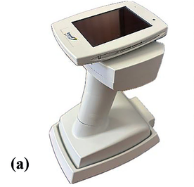
SpectroShade Micro
SpectroShade Micro is an imaging spectrophotometer. Its use for measuring anterior teeth is documented in Liondev et al.’s study of dental students. That population study describes tooth-colour differences; it is not a head-to-head trial establishing the best purchasing choice or a restoration-success rate.
Rayplicker Handy and other camera-based workflows
Borea describes Rayplicker Handy as a spectrophotometer providing tooth-colour and translucency maps. Its support materials distinguish acquisition and care requirements by device. Mapping functions do not remove the need for correct positioning or an appropriate reference shade guide. Smartphone and intraoral-scanner colour functions likewise require evidence for the actual hardware, software, calibration and indication; a colour image alone is not proof of calibrated shade measurement.
Translate the record into a restoration
The laboratory needs more than a shade code. Communicate the substrate colour, available restorative space, desired regional opacity or translucency, surface character and the material under consideration. A product family’s shade name is not a guarantee that different materials, thicknesses and processing routes will look identical.
In an in-vitro study by Czigola et al., substrate, ceramic thickness, translucency and try-in paste shade all affected the colour of A1 lithium-disilicate CAD/CAM crowns; try-in paste had the weakest effect among those tested. The work used a premolar model and specific materials. It supports considering the whole optical assembly, not a universal preparation thickness, a cement-only solution to a dark substrate, or an anterior clinical outcome guarantee.


Composite, glass-ceramic and zirconia options also differ in indication, processing, bonding and mechanical behaviour. Choose them through clinical assessment and the exact manufacturer’s instructions, rather than a colour-stability ranking assembled from unmatched studies. A highly translucent material may reveal the substrate; increasing opacity may help masking but can change the intended depth and appearance. Evaluate these trade-offs in the actual planned restoration.
Interpret colour differences without turning them into guarantees
Perceptibility and acceptability are different color-matching judgments. In a multicenter observer study of dental ceramic specimens under simulated clinical conditions, Paravina et al. (2015) reported 50:50% perceptibility and acceptability thresholds of ΔEab = 1.2 and 2.7, respectively, using CIELAB, and ΔE00 = 0.8 and 1.8 using CIEDE2000. These formula-specific experimental values should be interpreted in the study context rather than as universal cutoffs for individual patients.
Always name the formula beside a ΔE value. A perceptibility threshold concerns noticing a difference; an acceptability threshold concerns tolerating it. Neither measures restoration survival, comfort or overall satisfaction. The appropriate interpretation also depends on the specimen or tooth region, illumination and observers. Use instrumental results to inform a clinical discussion and visual comparison, not to overrule the patient’s experience.
Work through difficult matches
The following are hypothetical decision examples, not reports of treated patients or documented outcomes.
A single central incisor with a cervical-to-incisal gradient
First ask whether the neighbouring tooth has a consistent body colour or distinct regional features. Record the target tooth before dehydration and map what needs to be communicated: lightness, cervical chroma, incisal translucency, texture and any local characterisation. If an instrument and visual assessment disagree, repeat the acquisition under controlled conditions before deciding which result better represents the clinical target.
A discoloured substrate beneath a proposed restoration
Ask whether the planned material and available space can provide the required masking without compromising the clinical indication. Communicate the substrate with the laboratory and evaluate the proposed optical combination. Do not assume a more opaque cement will correct every mismatch or select a material from a stock image. The cited laboratory research explains why the layers interact; it does not prescribe treatment for this hypothetical patient.
At review, separate the questions: Is the regional colour close enough? Do contour, texture and gloss change the visual impression? Is the patient comfortable with the appearance? If the result is unacceptable, identify the likely contributor and reassess before an irreversible treatment step. The clinician remains responsible for the treatment decision.
Where AI fits
Artificial Intelligence in Shade Matching: Machine learning algorithms are being developed to analyze tooth color patterns and predict optimal shade distributions across restorations. A 2024 systematic review by Shetty et al. included 15 studies and concluded that lighting, the measuring device, the color-space model and the AI method affect shade-prediction accuracy.
The review concerns shade-prediction methods and their inputs. It does not establish that a particular current commercial tool will produce a satisfactory restoration in every patient. Evaluate the intended use, validation population, acquisition conditions and external testing before relying on a prediction. A visual preview also remains distinct from the optical behaviour of a manufactured restoration.
Practical takeaway
Build a shade record that another clinician or technician can understand and repeat. Preserve the measurement conditions, describe regional tooth characteristics, communicate the substrate and restorative material, and check the completed restoration visually as well as instrumentally where appropriate. Digital tools can support that process, while evidence limits and patient-specific assessment remain part of every decision.
References and source notes
- Sikri VK. Color: Implications in dentistry. J Conserv Dent. 2010;13(4):249–255. doi:10.4103/0972-0707.73381. Background review.
- Alamé C, Mehanna Zogheib C. The Effect of Dehydration on Tooth Color: A Prospective In Vivo Study. Cureus. 2023;15(11):e48140. PubMed.
- Liberato WF, Barreto IC, Costa PP, et al. A comparison between visual, intraoral scanner, and spectrophotometer shade matching: A clinical study. J Prosthet Dent. 2019;121(2):271–275. doi:10.1016/j.prosdent.2018.05.004.
- Dozić A, Kleverlaan CJ, El-Zohairy A, Feilzer AJ, Khashayar G. Performance of five commercially available tooth color-measuring devices. J Prosthodont. 2007;16(2):93–100. doi:10.1111/j.1532-849X.2007.00163.x.
- Paul SJ, Peter A, Rodoni L, Pietrobon N. Conventional visual vs spectrophotometric shade taking for porcelain-fused-to-metal crowns: a clinical comparison. Int J Periodontics Restorative Dent. 2004;24(3):222–231. PubMed.
- Czigola A, Abram E, Kovacs ZI, et al. Effects of substrate, ceramic thickness, translucency, and cement shade on the color of CAD/CAM lithium-disilicate crowns. J Esthet Restor Dent. 2019;31:457–464. doi:10.1111/jerd.12470. Laboratory study.
- Paravina RD, Ghinea R, Herrera LJ, et al. Color difference thresholds in dentistry. J Esthet Restor Dent. 2015;27 Suppl 1:S1-S9. doi:10.1111/jerd.12149. Publisher abstract
- Shetty S, Gali S, Augustine D, et al. Artificial intelligence systems in dental shade-matching: A systematic review. J Prosthodont. 2024;33(6):519-532. doi:10.1111/jopr.13805. Publisher abstract
- Liondev I, Dzhondrova I, Radev R, et al. Color Characteristics of the Anterior Teeth in Dental Students. J of IMAB. 2025;31(3):6322–6326. Article and SpectroShade figure source.
- Ciocan Pendefunda AA, Dascalu CG, Bahrim S, et al. Practical Implications in Contemporary Dental Aesthetics—Shade Selection Assessment Using Intraoral Scanners. Dent J. 2025;13(1):43. Easyshade figure source. Laboratory study; the figure is used for device identification.
Manufacturer links in the device section describe functions and instructions. They are not independent comparative-effectiveness evidence.

WHERE DIGITAL MEETS CLINICAL
Perio-Implantologist
International Digital Dentistry Speaker
Digital Occlusion Consultant
Founder of DDS
Founder of Occlusa AI Platform
 h.sharshar@ddschoology.com
About Dr. Haitham Sharshar
Dr. Haitham Sharshar is an international digital dentistry speaker, educator, and digital occlusion consultant based in Cairo, Egypt. His work focuses on functional digital dentistry, CAD/CAM, implantology, jaw-motion analysis, and the integration of digital diagnostics into clinical treatment planning.
As Founder and Scientific Coordinator of Digital Dentistry Schoology (DDS), he has trained more than 3,270 dentists and dental technicians through university programs, international conferences, and hands-on courses. His teaching connects patient-specific diagnostic records with digital design and clinical workflows, helping clinicians and technicians understand how function and occlusion inform restorative planning.
Dr. Sharshar is a certified trainer for zebris JMA-Optic+ jaw-motion analysis and MyoWise dental EMG. His educational and consulting work brings together patient diagnostics, jaw-motion records, muscle-activity data, digital occlusion, and AI-supported workflows.
He is Founder and Clinical Director of Occlusa, an AI-supported platform for organizing clinical information and supporting clinician-led review and treatment planning. He also owns HS Dental Clinic in Cairo, where his clinical focus includes full-mouth digital rehabilitation and smile design.
His speaking and training topics include functional digital dentistry, digital occlusion, jaw-motion analysis, CAD/CAM workflows, digital implantology, and the practical integration of AI-supported tools into dental education and clinical practice.
Happy to collaborate on spreading digital dentistry.