
Bone Grafting Materials: Comparing Evidence for Oral Reconstruction
Which bone graft fits the oral reconstruction you are planning? Begin with the defect and intended restoration, then ask what evidence the exact material has in that setting.
Autogenous bone, donated human tissue, animal-derived scaffolds and synthetic substitutes differ in composition and processing. The studies discussed here do not establish one graft class as best for every patient. (Troeltzsch et al., 2016; Urban et al., 2019; Khan et al., 2005)
This overview connects graft biology with evidence from extraction sockets, sinus augmentation and ridge reconstruction. It then follows one published anterior-maxillary case through digital planning, augmentation and restoration. Biopsy composition, ridge dimensions and implant survival remain separate outcomes. This is a selected-evidence teaching article, not a systematic review or a report of a DDS-treated patient. (Danesh-Sani et al., 2017; Starch-Jensen et al., 2018; Tallarico et al., 2020)
What do the biological terms mean?
A useful comparison starts by separating a scaffold, a biological signal and a living cell. Those functions may coexist, but naming them does not establish that a graft will achieve a particular result in a patient. The host bed and handling of the graft also affect incorporation. (Albrektsson and Johansson, 2001; Khan et al., 2005)
Cells, signals and scaffold are different contributions
Osteogenesis means new bone formation by bone-forming cells. Autogenous grafts can provide viable cells as well as matrix and scaffold. Conventional acellular substitutes differ from cellular allograft products, some of which contain living cells; the presence of cells alone is not evidence of better clinical outcomes. (Khan et al., 2005; CDC, 2023 outbreak)
Osteoinduction concerns signals that recruit cells toward a bone-forming lineage. Osteoconduction concerns a surface or scaffold along which bone can grow. These are explanatory properties, not a material league table. For example, the induction observed with a particular demineralized allograft preparation cannot be assumed for every preparation. (Albrektsson and Johansson, 2001; Schwartz et al., 1996)
Osseointegration describes stable implant anchorage through direct contact between living bone and the implant. Keep it distinct from graft incorporation and remodeling: the persistence of particles, the tissue seen in a biopsy and the performance of an implant answer different questions. (Albrektsson and Johansson, 2001; Danesh-Sani et al., 2017; Starch-Jensen et al., 2018)
What does the graft category tell us?
Autogenous bone: the donor-site trade-off
Autogenous bone is harvested from the patient. It can bring viable bone-forming cells, matrix-associated signals and an osteoconductive scaffold, but the tissue collected and its handling matter. It avoids a separate human-donor transmission pathway while adding a harvesting procedure; that does not remove surgical infection or other procedural risks. (Khan et al., 2005; Misch, 1997)
Misch’s clinical study followed 50 patients receiving mandibular symphysis or ramus grafts before implant placement. The symphysis provided corticocancellous blocks and the ramus predominantly cortical grafts. Donor-site choice was related to the required graft and access to the recipient site; the study also reported differing donor-site complications. It does not compare all skeletal donor sites. (Misch, 1997)
A compromised recipient blood supply is not, by itself, a reason to select a nonvascularized autograft. Graft incorporation depends on the host and graft environment, including vascularity and mechanical conditions. Large load-bearing reconstruction and vascularized graft transfer are separate decisions that this oral-augmentation overview does not resolve. (Khan et al., 2005)
Allografts: preparation and safety
Allografts are donated human tissue. Donor screening, testing and processing reduce infectious-disease transmission risk but do not eliminate it. Processing names are not interchangeable performance claims: the LifeNet dental allograft instructions distinguish frozen, freeze-dried and Preservon-preserved forms, and warn about possible disease transmission and rejection or allergic reactions. (FDA tissue safety; LifeNet Health IFU)
Demineralized freeze-dried bone allograft (DFDBA) may show osteoinductive activity, but Schwartz and colleagues found substantial variation among preparations in an animal assay. That finding does not verify a current batch’s human clinical effect. Nor does the label “freeze-dried” establish preserved mechanical performance, faster incorporation or a lower price than another graft. (Schwartz et al., 1996; LifeNet Health IFU)
The CDC documented a 2023 tuberculosis outbreak associated with a bone-allograft product containing live cells. This establishes that donor-derived transmission can occur; it does not supply a risk estimate for every processed dental allograft. Distinguish cellular products from conventional processed acellular preparations when discussing both biological claims and safety. (CDC, 2023 outbreak)
In a six-month randomized study of adults requiring nonmolar extraction and delayed implant placement, Iasella and colleagues evaluated tetracycline-hydrated freeze-dried bone allograft with a collagen membrane against extraction alone. The studied protocol limited dimensional loss, but it tested a combined intervention. It does not establish the suitability of every allograft for every dental or orthopedic defect. (Iasella et al., 2003)
A donated graft avoids harvesting that component from the patient. Identify the exact preparation, supply and applicable product instructions before discussing that trade-off. For example, the dental-indication list in the LifeNet document reviewed here is explicitly marked for Mexico; it cannot be used as a worldwide indication list. (LifeNet Health IFU)
Xenografts: origin and processing
A xenograft is derived from another species. For a named example, Geistlich’s Australian patient information describes Bio-Oss as a bovine-derived osteoconductive scaffold that resorbs slowly over many years. That product description is not a resorption timetable for all animal-derived grafts, nor a comparative clinical advantage. (Geistlich patient leaflet, 2025)
Processing can change what remains in an animal-derived graft. Nannmark and Sennerby studied collagenated porcine particles, with and without added collagen gel, in rabbit maxillary defects. They observed osteoconduction and resorption in that model. The experiment does not establish that porcine materials more closely resemble human bone or outperform other grafts in people. (Nannmark and Sennerby, 2008)
A clinical example is Urban and colleagues’ horizontal-augmentation series using autogenous particles plus anorganic bovine bone mineral under a resorbable collagen membrane. It supports a report of that combined approach. It does not justify a class-wide list covering every periodontal, cystic, sinus or ridge defect, or show how much of the result came from either graft component. (Urban et al., 2013)
As an educational planning step, make the material’s human, animal or synthetic origin explicit when discussing options with the patient. The source category should be known before asking about acceptance of donor tissue or an additional harvesting procedure. A category label alone does not identify the full composition of a composite product. (Geistlich patient leaflet, 2025; LifeNet Health IFU)
Synthetic substitutes: formulation and evidence
The studies and device records below concern distinct synthetic formulations, including calcium-phosphate ceramics, calcium sulfate and a named bioactive glass. They are not a single interchangeable class. A fully synthetic formulation avoids a donor-tissue transmission pathway, but contamination and surgical infection remain possible; check whether a composite also contains donor-derived components. (Yuan et al., 2010; De Leonardis and Pecora, 2000; FDA PerioGlas clearance)
Yuan and colleagues demonstrated osteoinduction by selected engineered calcium-phosphate ceramics in laboratory and animal models without added growth factors. This prevents a blanket statement that no synthetic material can be osteoinductive. It also does not establish that every marketed calcium-phosphate graft has that property or is superior in human dental care. (Yuan et al., 2010)
In a prospective sinus-augmentation study, De Leonardis and Pecora found that calcium-sulfate application technique affected shrinkage and resorption. Particles produced by its resorption were observed in six-month test-group specimens and were no longer detected at nine months. These were not described as unchanged calcium sulfate; material disappearance also does not prove complete replacement by mature bone. (De Leonardis and Pecora, 2000)
For a product-specific example, the US PerioGlas clearance record describes a particulate calcium-phosphosilicate (45S5 Bioglass) non-structural scaffold with specified dental and craniofacial uses, including intrabony periodontal defects, extraction sites and sinus lifts. This historical device record is not a current worldwide instruction sheet or evidence that all bioactive glasses have those indications. Its cell-culture language does not establish a superior clinical outcome. (FDA PerioGlas clearance)
“Composite” and “surface-modified” describe material construction, not a proven benefit. Kim, Yang and Lee studied atelocollagen-precoated biphasic calcium-phosphate granules in cell experiments and rabbit calvarial defects. Their 2017 paper concerns that coating; it does not test an RGD-peptide intervention or establish human dental efficacy. (Kim et al., 2017)
Which outcomes are comparable?
A comparison is useful only when its endpoint matches the clinical question. Socket dimensions address ridge contraction after extraction; a sinus biopsy describes sampled tissue; implant survival follows a different event over a specified interval. The studies below therefore should not be collapsed into one ranking. (Avila-Ortiz et al., 2014; Danesh-Sani et al., 2017; Starch-Jensen et al., 2018)
Extraction sockets: dimensional preservation is the measured benefit
Vignoletti and colleagues found reduced horizontal and vertical contraction with ridge-preservation therapies, without establishing a preferred biomaterial or long-term implant benefit. Avila-Ortiz and colleagues’ 2014 review concerned adult nonmolar extraction sockets. Its graft comparisons and exploratory subgroups do not establish an allograft–xenograft mixture as superior. (Vignoletti et al., 2012; Avila-Ortiz et al., 2014)
In Iocca and colleagues’ small network of six studies, freeze-dried bone graft plus membrane ranked most favorably for height preservation and autologous bone marrow for width preservation. Those were indirect, endpoint-specific rankings with evidence limitations. They do not show that one graft class universally outperforms another. (Iocca et al., 2017)
Sinus augmentation: separate histology from implant survival
Danesh-Sani and colleagues synthesized histomorphometric findings from 136 human studies. Autogenous bone had the highest new-bone fraction in that synthesis, and healing time influenced the measurements. The paper appeared online in 2016 and in the 2017 journal issue. A tissue fraction describes composition; it is not a rate of bone growth or an implant-survival result. (Danesh-Sani et al., 2017)
Starch-Jensen and colleagues’ 2018 review reported high long-term implant survival across the included sinus-augmentation approaches, but identified no long-term randomized comparison of grafting modalities. This cannot establish that the material with the largest biopsy new-bone fraction also produces the best long-term implant outcome. (Starch-Jensen et al., 2018; Danesh-Sani et al., 2017)
Horizontal augmentation: keep the observed gain in context
Troeltzsch and colleagues reported a weighted mean horizontal gain of 3.7 ± 1.2 mm (mean ± SD) for particulate grafts across heterogeneous studies; block grafts achieved approximately 1 mm more. These pooled observations are not a prediction for an individual defect and do not establish universal superiority of autogenous blocks. (Troeltzsch et al., 2016)
Urban and colleagues’ 2013 prospective case series used a 1:1 mixture of particulate autogenous bone and anorganic bovine bone mineral with a resorbable collagen membrane for horizontal augmentation. The ratio and membrane are part of that reported protocol. The uncontrolled series does not establish an optimal mixture or isolate each component’s contribution. (Urban et al., 2013)
Vertical augmentation: technique and complications remain visible
Rocchietta and colleagues’ review emphasized limitations in the generalizability of vertical-augmentation evidence. Urban and colleagues’ 2019 synthesis found procedure-dependent bone gain and common complications. The latter is a review of several procedures, not a single trial proving a particular composite graft to be the most predictable. (Rocchietta et al., 2008; Urban et al., 2019)
What changes when materials are combined?
Combining materials may be a rationale worth examining, but the mixture itself needs evidence. The 1:1 mixture in Urban’s horizontal-augmentation series and the mixture in the published case below belong to their respective protocols. Neither establishes a universal recipe. The sources reviewed here also do not substantiate an allograft–xenograft superiority claim or a specific layering pattern that guarantees vascularization. (Urban et al., 2013; Avila-Ortiz et al., 2014; Tallarico et al., 2020)
Platelet preparations: which endpoint?
Platelet-rich plasma (PRP) and platelet-rich fibrin (PRF) are different preparations. For periodontal intrabony defects, Del Fabbro and colleagues’ 2011 review found an adjunctive PRP benefit for clinical attachment level with graft materials when guided tissue regeneration (GTR) was not used; no adjunctive PRP effect on that endpoint was found when GTR was used. This does not establish PRF equivalence or improved early healing in every grafted site. (Del Fabbro et al., 2011)
BMPs: product, carrier and indication
In the United States, INFUSE Bone Graft combines rhBMP-2 with its absorbable collagen sponge. The dental approval is as an autograft alternative for sinus augmentation and localized alveolar ridge defects associated with extraction sockets. This is a specified protein-and-carrier system, not a general endorsement of any BMP preparation or carrier. (FDA dental approval; Medtronic dental safety summary)
The manufacturer’s US dental safety summary lists hypersensitivity to rhBMP-2, bovine type I collagen or other formulation components, pregnancy and active infection at the operative site as contraindications. It also says not to use the product near an existing or resected tumor, in patients with active malignancy or during malignancy treatment. Skeletally immature patients have not been studied. (Medtronic dental safety summary)
The original 2007 dental label describes swelling and the potential for unwanted bone formation. That historical label and the manufacturer’s brief safety summary do not replace the complete current instructions applicable to the product and country. No dose, off-label use or patient-specific treatment protocol is proposed here. (FDA dental label, 2007; Medtronic dental safety summary)
A separate US INFUSE tibial indication concerns skeletally mature patients with acute open tibial shaft fractures stabilized with an intramedullary nail after appropriate wound management; application must occur within 14 days after the initial fracture. It is not a general nonunion indication and cannot be transferred to dental defects. (FDA tibial indication)
Historically, OP-1 Implant (rhBMP-7) received a humanitarian device exemption for recalcitrant long-bone nonunions after alternatives had failed and autografting was not feasible. Its label states that effectiveness for that use had not been demonstrated. This differs from the dental INFUSE approval; these records do not establish current OP-1 availability or authorization outside the United States. (FDA OP-1 HDE; OP-1 package insert)
Custom scaffolds: fit and regeneration
Rasperini and colleagues’ 2015 report described a digitally designed and printed scaffold in one human periodontal case. Initial coverage was followed by exposure and removal around 14 months, with limited bone repair. The report illustrates an early clinical translation attempt; it does not show that anatomical fit, designed pores or printability alone ensure durable regeneration. (Rasperini et al., 2015)
Does this evidence fit the defect?
The comparison below links the defect, graft, measured outcome and follow-up. It helps judge how far a study or product instruction applies to the planned reconstruction; it does not select a graft or replace clinical assessment. (Khan et al., 2005; Troeltzsch et al., 2016; Urban et al., 2019)
Does the source answer this reconstruction question?
- Which defect was studied?
- Setting
- Troeltzsch and colleagues reviewed 184 ridge-augmentation papers involving 6,182 patients.
- Finding
- Weighted mean horizontal gain with particulate grafts was 3.7 ± 1.2 mm (mean ± SD).
- Follow-up
- The weighted mean observation period was 27.4 months; individual studies ranged from 3 to 168 months.
- Limit
- This pooled ridge gain is not an individual forecast, a sinus-biopsy result or proof that one particulate class is superior.
- What does the donor-site comparison show?
- Setting
- Misch studied 50 patients receiving mandibular symphysis or ramus onlay grafts before implant placement.
- Finding
- Ramus harvesting was associated with fewer donor-site complications in this cohort.
- Timing
- Implants were placed after 4–6 months of graft healing; this interval is not a long-term survival endpoint.
- Limit
- A donor-site comparison does not establish a graft indication for a poorly vascularized recipient bed. Graft incorporation also depends on the host and local conditions.
- Which formulation is actually covered?
- Setting
- The cited sources are LifeNet’s dental allograft instructions, the PerioGlas clearance record and Medtronic’s dental safety summary.
- What they establish
- Their statements concern the named product, preparation and indicated use.
- Outcome and follow-up
- These product documents are not a shared head-to-head comparison with one clinical endpoint and follow-up period.
- Limit
- An indication for one formulation does not demonstrate equivalence across a graft class.
- What was measured, and for how long?
Sinus tissue composition, ridge dimensions and implant survival answer different questions. (Danesh-Sani et al., 2017; Starch-Jensen et al., 2018; Troeltzsch et al., 2016; Urban et al., 2019)
- Case setting
- Tallarico’s single-patient report combined a customized titanium mesh, autogenous and bovine bone, and several adjuncts in an anterior-maxillary horizontal defect.
- Observed outcome
- Treatment progressed through implant placement and delivery of the definitive restoration.
- Two follow-up clocks
- The review was one year after implant placement, but only two months after delivery of the definitive restoration.
- Limit
- The report does not isolate a graft-component effect, measure graft histology or establish long-term definitive-restoration survival.
The clinical sequence below shows how the authors connected augmentation, implant planning and restoration in one patient. Their treatment choices and observed result remain separate from a general material recommendation.
One published case: from a horizontal defect to a digital restoration
This external single-patient report shows how graft selection, prosthetic planning and a custom titanium mesh were combined in the anterior maxilla. It illustrates the authors’ decisions; it does not establish a superior graft material or a treatment protocol for every patient.
The source reports written consent for publication of clinical and radiologic data. The sequence below uses intraoral and digital records; it does not identify the patient or imply treatment by DDS.
Use the buttons or swipe horizontally. With the steps focused, use Left and Right Arrow, Home or End. You can also read all steps as a vertical list.
1. Clinical baseline
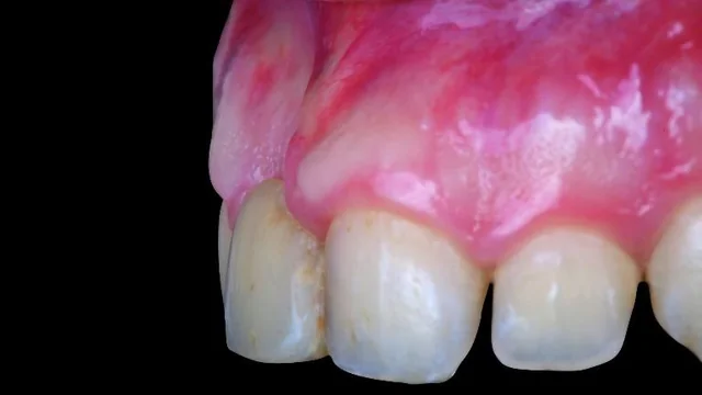
The adult patient had a missing maxillary right central incisor and a horizontal ridge deficiency following earlier trauma. Source, Figure 1. Teaching point: Relate the prosthetic complaint to the tissue contour; this photograph does not measure the defect.
Interpretation limit: A published case from an external clinical team; not a DDS-treated patient.
2. CBCT and prosthetic planning
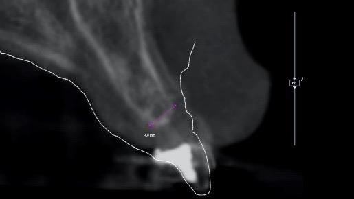
CBCT and intraoral-scan data informed prosthetically driven planning. The paper reports about 4 mm of crestal width and no vertical defect. Source, Figure 2. Open Figure 2 at its largest reviewed size (515 × 290; new tab).
Teaching point: The clinical question was horizontal augmentation in this patient; the image is not a universal width threshold.
Interpretation limit: The source figure is only 515 × 290 pixels; the reported measurement is also given as readable text.
3. Designing the titanium mesh
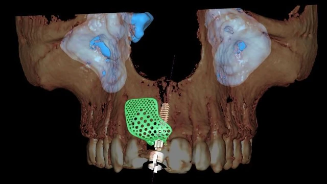
A CAD designer shaped the custom titanium mesh using the contralateral anterior-maxillary outline and the planned implant position. Source, Figure 3. Teaching point: This illustrates how the prosthetic plan and intended contour informed space maintenance.
Interpretation limit: The article does not provide a downloadable CAD file or validated design tolerances.
4. Manufacturing the mesh and guide
Documented text-only stage: the custom titanium mesh and surgical template were manufactured and shown on printed models in the publication. Source, Figure 4 and page 4.
Teaching point: connect the virtual mesh design to the physical parts used during treatment.
Interpretation limit: the source shows the finished parts; it does not provide a live printing-process sequence or downloadable CAD files. The model photograph is not reproduced in this adaptation.
5. Implant placement and augmentation
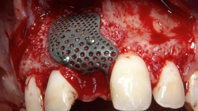
The report describes guided implant placement, local autogenous-bone collection and a 1:1 mixture of autogenous bone and anorganic bovine bone (Bio-Oss), followed by mesh fixation with two pins. Source, Figure 5. Teaching point: Connect graft selection to the reported defect and space-maintaining scaffold.
Interpretation limit: The mixture is documented in the text; this photograph does not independently establish its composition. One case cannot identify a superior graft or universal protocol.
6. Four-month healing review
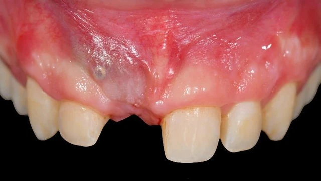
The authors report an uncomplicated four-month healing period before second-stage surgery. Source, Figure 7. Teaching point: Distinguish an observed healing review from proof of graft histology.
Interpretation limit: The image does not quantify vital bone, residual graft or ridge-volume gain.
7. Mesh removal and re-entry
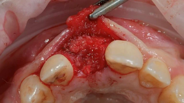
At four months, second-stage surgery included mesh removal and soft-tissue management. The text then describes PRF application and a screw-retained provisional restoration. Source, Figure 8. Teaching point: Link the provisional-restoration phase to the earlier augmentation rather than skipping directly to the final crown.
Interpretation limit: PRF use is a reported adjunct; this uncontrolled case cannot isolate its effect. A separate image of PRF preparation is not supplied.
8. Capturing the restorative position
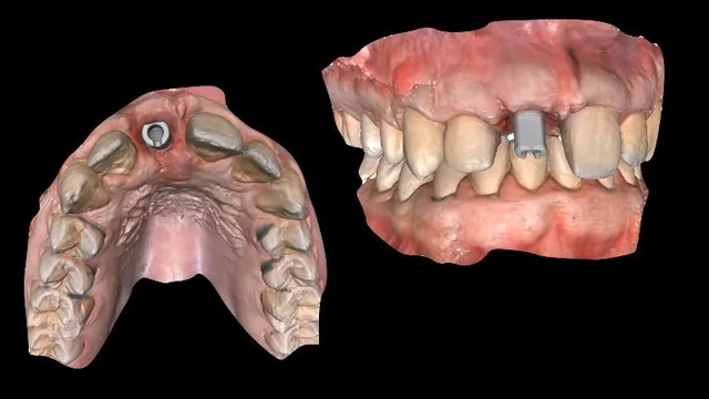
Three months after second-stage surgery, the adjacent central incisor was prepared for a veneer and the definitive digital impression recorded the implant position using a scan body. Source, Figure 11. Teaching point: Show how clinical tissues and the implant position entered the restorative digital workflow.
Interpretation limit: The source does not supply a quantitative scan-accuracy assessment for this patient.
9. Restoration design
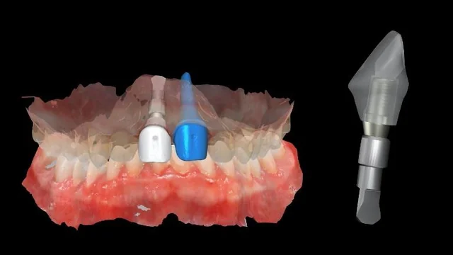
The restorative workflow used digital design and printed models with a digital implant analog and a removable die at the natural-tooth position. Source, Figure 12. Teaching point: Explain the connection between the scan record and laboratory design.
Interpretation limit: Source caption uses “removable abutment”; the teaching text uses “removable die” to distinguish the natural-tooth model component from an implant abutment.
10. Manufactured definitive restoration
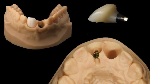
The definitive implant crown was zirconia layered with feldspathic ceramic and bonded extraorally to a titanium abutment. A ceramic veneer was also delivered to the adjacent incisor. Source, Figure 13. Teaching point: Keep the actual restorative construction visible when interpreting the aesthetic result.
Interpretation limit: Do not call this hybrid crown wholly metal-free: the case includes a titanium abutment.
11. Reported clinical endpoint
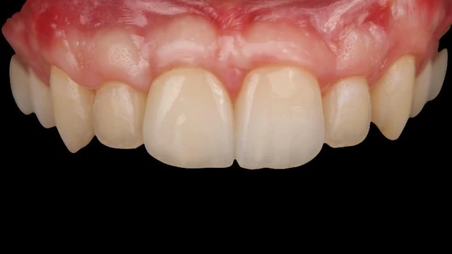
This is the reported one-year review after implant placement, two months after definitive-restoration delivery. Source, Figure 15. Teaching point: Keep the implant-placement and restoration-delivery clocks separate.
Interpretation limit: A single short follow-up case does not establish long-term survival or comparative efficacy.
12. Radiographic sequence
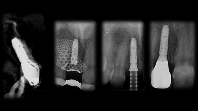
Source-sequence overview: the publication reports radiographic follow-up through one year after implant placement. This small composite is not presented for radiographic measurement or diagnosis. Source, Figure 17. Open Figure 17 at its largest reviewed size (640 × 360; new tab).
Teaching point: Pair the visible clinical result with the authors’ radiographic follow-up.
Interpretation limit: The caption does not assign exact dates to each of the four panels; do not invent them. These images do not establish histology or quantified bone gain.
Case and image source: Marco Tallarico, Chang-Joo Park, Aurea Immacolata Lumbau, Marco Annucci, Edoardo Baldoni, Alba Koshovari and Silvio Mario Meloni. Customized 3D-Printed Titanium Mesh Developed to Regenerate a Complex Bone Defect in the Aesthetic Zone: A Case Report Approached with a Fully Digital Workflow. Materials. 2020;13(17):3874. © 2020 the authors; licensee MDPI. Reused under CC BY 4.0.
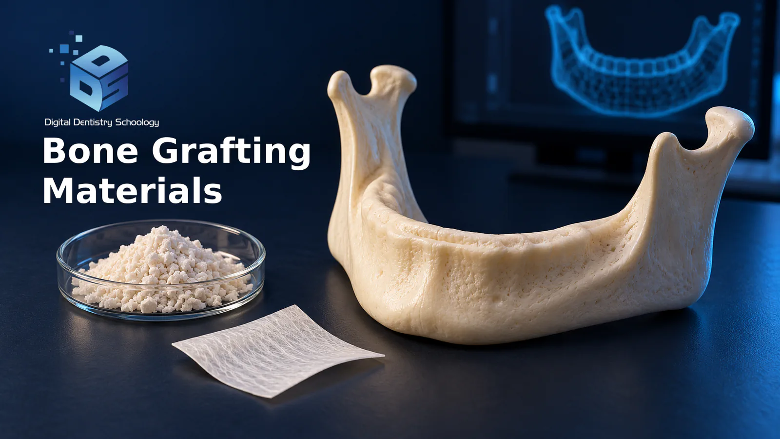
Case figures: 1–3, 5, 7–8, 11–13, 15 and 17; the manufacturing account refers to Figure 4 and page 4. AI-generated illustration of bone-graft materials and digital planning; not a patient image or treatment plan.
Display images resized and encoded as WebP; Figure 2 has unnecessary timestamp metadata removed. Source photographs and anatomy have not been generated, retouched or relabeled. Captions and teaching commentary are adapted for DDS. No endorsement by the source authors is implied.
Follow-up boundary: one year after implant placement was only two months after delivery of the definitive restoration. The authors report favorable clinical and radiographic findings in this case; the record does not compare grafts or establish long-term outcomes. The paper acknowledges donated mesh, models and templates from New Ancorvis.
What matters for the dentist and laboratory?
Before applying a finding, ask whether its defect, procedure, product and follow-up match the case in front of you. The points below support discussion between the dentist and laboratory; they do not provide a patient-specific grafting or manufacturing protocol. (Troeltzsch et al., 2016; Urban et al., 2019; Tallarico et al., 2020)
For the dentist
- State the endpoint before comparing materials. A biopsy fraction does not answer an implant-survival question. Record the defect, intended procedure, material, measured outcome and follow-up alongside any number used in the discussion; pooled gains are not individual forecasts. (Danesh-Sani et al., 2017; Starch-Jensen et al., 2018; Troeltzsch et al., 2016)
- Include the source and donor trade-off. Autogenous harvesting adds a donor-site decision; allograft screening does not eliminate transmission risk. Identify the exact tissue preparation and relevant product instructions before discussing risks or assuming equivalence. The cited sources do not provide a universal comparative infection rate. (Misch, 1997; FDA tissue safety; LifeNet Health IFU)
- Keep a case’s limits beside its outcome. The Tallarico report used a combined graft and several adjuncts. Document its two follow-up clocks: one year after implant placement and only two months after definitive-restoration delivery. The case cannot identify which component caused the outcome or prove long-term survival. (Tallarico et al., 2020)
For the laboratory and digital-planning team
- Connect the prescribed contour to the supplied records. In the published case, the implant plan and contralateral maxillary outline informed the mesh design. Keep that relationship traceable in discussion with the clinician; the paper supplies no downloadable CAD file or validated universal design tolerances. (Tallarico et al., 2020)
- Name the actual restoration and what was checked. The case’s definitive implant crown included zirconia, feldspathic layering and a titanium abutment. Keep the scan, design and manufactured construction distinguishable when explaining the result; a visually successful restoration is not a measurement of graft histology or comparative material efficacy. (Tallarico et al., 2020)
For oral reconstruction, the most useful comparison is specific: this material, in this defect, with this procedure, measured at this interval. The biological categories help organize the question; studies, product instructions and the recipient environment determine how far an answer can go. A clear comparison preserves those limits while showing the clinical and digital steps that connect the graft to the eventual restoration. (Khan et al., 2005; Troeltzsch et al., 2016; Urban et al., 2019; Tallarico et al., 2020)
Sources and scope
Study findings, device records and manufacturer information serve different purposes. The links below map to the passages above; the published case retains its own full image credit and licence notice.
- FDA: Tissue and Tissue Product Questions and Answers. Official US regulator information.
- CDC: Second Nationwide Tuberculosis Outbreak Caused by Bone Allografts Containing Live Cells—United States, 2023. Primary outbreak investigation / MMWR.
- FDA PMA P050053: INFUSE Bone Graft dental approval. Official US PMA record.
- Medtronic current US dental INFUSE indications/safety brief. Official manufacturer US labeling summary.
- FDA original dental INFUSE physician labeling (2007). Original US FDA physician labeling, M704819B001 Rev A, 8 March 2007.
- FDA HDE H010002: OP-1 Implant. Official US humanitarian device exemption record.
- FDA original OP-1 Implant package insert. Original humanitarian-device package insert.
- FDA PMA P000054: INFUSE tibial trauma indication. Official US PMA record.
- Albrektsson and Johansson (2001), Osteoinduction, osteoconduction and osseointegration. Conceptual scientific article; not a comparative clinical trial.
- Schwartz et al. (1996), Ability of commercial DFDBA to induce new bone formation. Primary preclinical ectopic implantation experiment.
- Yuan et al. (2010), Osteoinductive ceramics as a synthetic alternative to autologous bone grafting. Primary in-vitro and animal experiments.
- Craig M. Misch (1997), Comparison of intraoral donor sites for onlay grafting prior to implant placement. Primary comparative clinical study.
- Vignoletti et al. (2012), Surgical protocols for ridge preservation after tooth extraction. Original systematic review, retrieved directly.
- Iocca, Farcomeni, Pardiñas Lopez and Talib (2017), Alveolar ridge preservation: Bayesian network meta-analysis. Original systematic review / network meta-analysis of RCTs.
- Danesh-Sani, Engebretson and Janal (2017; online 2016), Histomorphometric results after sinus floor augmentation. Original systematic review / meta-analysis.
- Starch-Jensen et al. (2018), Long-term studies of maxillary sinus floor augmentation. Original systematic review / meta-analysis.
- Troeltzsch et al. (2016), Clinical efficacy of grafting materials in alveolar ridge augmentation. Original systematic review.
- Urban et al. (2013), Horizontal ridge augmentation with collagen membrane and autogenous bone/ABBM. Primary prospective uncontrolled case series.
- Rocchietta, Fontana and Simion (2008), Clinical outcomes of vertical bone augmentation. Original systematic review.
- Urban, Montero, Monje and Sanz-Sánchez (2019), Effectiveness of vertical ridge augmentation interventions. Original systematic review / meta-analysis.
- Nannmark and Sennerby (2008), Porcine grafts in rabbit maxillary defects. Primary animal experiment.
- De Leonardis and Pecora (2000), Calcium sulfate sinus augmentation histology. Primary prospective clinical study; two application-technique groups.
- Del Fabbro et al. (2011), Platelet concentrate for surgical periodontal treatment. Original systematic review / meta-analysis of RCTs.
- Kim, Yang and Lee (2017), Atelocollagen-precoated BCP. Primary in-vitro and rabbit-calvarial study.
- Avila-Ortiz et al. (2014), Effect of alveolar ridge preservation after tooth extraction. Original systematic review / meta-analysis of RCTs.
- Rasperini et al. (2015), 3D-printed bioresorbable scaffold for periodontal repair. Primary single-human case report with digital planning/fabrication.
- Geistlich Bio-Oss / Bio-Oss Pen patient information leaflet, Australia (2025). Official manufacturer patient information.
- Khan et al. (2005), The biology of bone grafting. Original scientific review; not a head-to-head clinical trial.
- Tallarico et al. (2020), Customized 3D-Printed Titanium Mesh Developed to Regenerate a Complex Bone Defect in the Aesthetic Zone: A Case Report Approached with a Fully Digital Workflow. Primary single-patient clinical case report with digital planning, fabrication and restorative stages.
- LifeNet Health: Sterile Dental Bone Allograft Bio-Implants, Instructions for Use, 63-0016-11 REV.01. Official manufacturer instructions.
- FDA 510(k) K053387: PerioGlas Bone Graft Particulate. Official US clearance record and manufacturer 510(k) summary; decision 2006-02-14.
- Iasella et al. (2003), Ridge preservation with freeze-dried bone allograft and a collagen membrane compared to extraction alone for implant site development. Primary randomized controlled blinded clinical/histologic study.

WHERE DIGITAL MEETS CLINICAL
DIGITAL DENTISTRY SCHOOLOGY
Perio-Implantologist
International Digital Dentistry Speaker
Digital Occlusion Consultant
Founder of DDS
Founder of Occlusa AI Platform
 h.sharshar@ddschoology.com
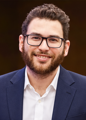
Written by Dr Haitham Sharshar
Dr. Haitham Sharshar is an international digital dentistry speaker, educator, and digital occlusion consultant based in Cairo, Egypt. His work focuses on functional digital dentistry, CAD/CAM, implantology, jaw-motion analysis, and the integration of digital diagnostics into clinical treatment planning.
As Founder and Scientific Coordinator of Digital Dentistry Schoology (DDS), he has trained more than 3,270 dentists and dental technicians through university programs, international conferences, and hands-on courses. His teaching connects patient-specific diagnostic records with digital design and clinical workflows, helping clinicians and technicians understand how function and occlusion inform restorative planning.
Dr. Sharshar is a certified trainer for zebris JMA-Optic+ jaw-motion analysis and MyoWise dental EMG. His educational and consulting work brings together patient diagnostics, jaw-motion records, muscle-activity data, digital occlusion, and AI-supported workflows.
He is Founder and Clinical Director of Occlusa, an AI-supported platform for organizing clinical information and supporting clinician-led review and treatment planning. He also owns HS Dental Clinic in Cairo, where his clinical focus includes full-mouth digital rehabilitation and smile design.
His speaking and training topics include functional digital dentistry, digital occlusion, jaw-motion analysis, CAD/CAM workflows, digital implantology, and the practical integration of AI-supported tools into dental education and clinical practice.
Happy to collaborate on spreading digital dentistry.