
A Technique for Gingival Displacement Before Digital Scanning
Soft-tissue management, evidence and limits for restorative digital impressions
Summary
Gingival displacement aims to make a preparation margin accessible for recording while limiting tissue injury. Cord, paste, foam and laser approaches should be judged by the clinical task and the outcomes actually measured. Greater displacement alone does not establish a more accurate restoration, better comfort or superior long-term tissue health. This overview separates a published technique citation, comparative clinical research and manufacturer product information.
The original technique and what this overview can establish
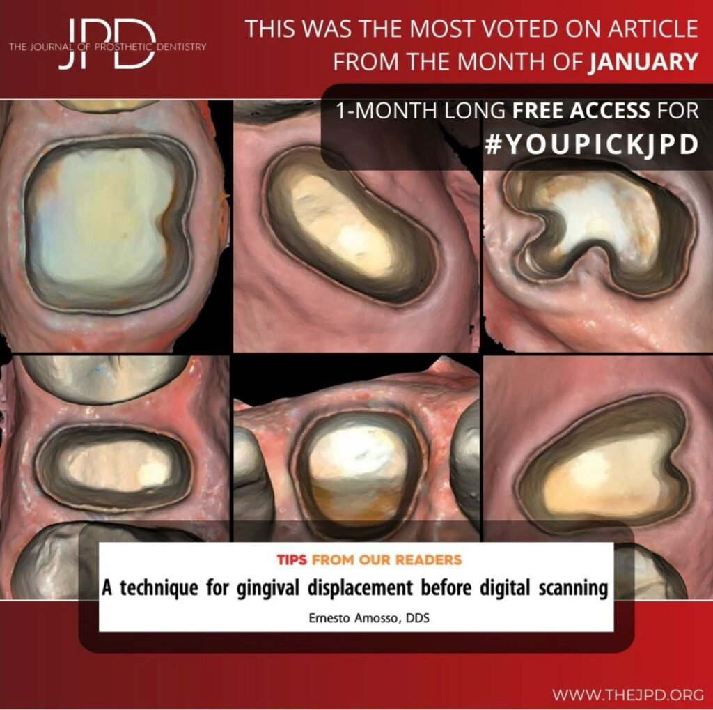
Ernesto Amosso authored A technique for gingival displacement before digital scanning in the Journal of Prosthetic Dentistry, published online in November 2024 and in the December 2025 issue. Its verified DOI is 10.1016/j.prosdent.2024.09.020. The bibliographic record has no abstract; the full text was unavailable for this review. Consequently, this overview does not reconstruct Amosso’s procedure, identify its materials or claim outcomes from it. Readers seeking that specific protocol should consult the original publication. [1]
Separate the clinical questions
- Margin access: Can the finish line be inspected and recorded?
- Field control: Are blood, fluid and residual material interfering with the record?
- Tissue response: What happens to gingival height and health after treatment?
- Restorative outcome: Does the completed restoration fit and function acceptably?
These questions are related, but answering one does not answer all four. A scan showing wider sulcular access is not itself a test of crown fit or survival. Equally, a product photograph cannot demonstrate atraumatic placement or patient comfort.
Comparative clinical evidence: displacement and tissue change
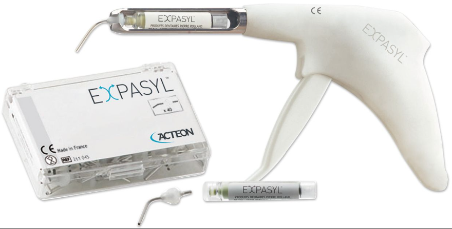
El Ashry and colleagues studied 32 participants requiring full crowns on maxillary premolars. The randomized groups used cord with astringent, Expasyl paste with astringent, Magic FoamCord without astringent, or laser troughing. The investigators measured displacement in digital impressions and gingival-height change through one month after crown cementation. Cord produced the greatest horizontal and vertical displacement, but also the greatest one-month gingival-height loss; Magic FoamCord followed cord for displacement. The authors cautioned against traumatic pressure with cord. [2]
This small, indication-specific trial supports weighing access against tissue effects. Its measured endpoints do not establish a universal winner for full-arch work, every periodontal condition, patient comfort or restoration longevity. The findings should not be converted into blanket “high” and “low” rankings for cord, paste and laser accuracy.
A separate randomized study of 40 participants examined double cord and aluminum-chloride paste before conventional impressions, comparing healthy gingiva with experimentally induced mild gingivitis. Cord’s advantage in sulcus representation was reduced with mild inflammation; marginal gingival-height change was not influenced by gingival condition in that study. It informs the importance of tissue condition, but its conventional-impression design should not be relabelled a digital crown-accuracy trial. [3]
Methods and product identity
Cord placement, material-assisted displacement and laser troughing are different interventions. A family name does not identify a formulation, device setting or protocol. Use the exact product’s current instructions and appropriate clinical training; this overview supplies no universal exposure time, packing force or laser setting.
| Example | What is identified | Interpretation limit |
|---|---|---|
| Expasyl / ACTEON | Retraction paste; the manufacturer also lists Expasyl Exact. [4] | Confirm the actual variant. Product descriptions do not establish comparative restoration accuracy or absence of injury. |
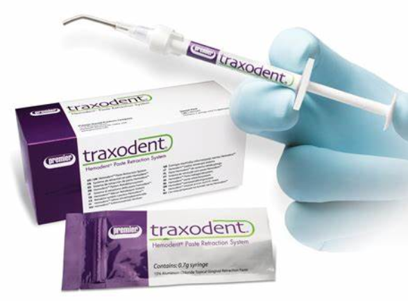
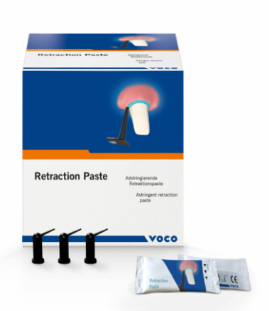
| Traxodent / Premier Dental | A hemostatic retraction paste system and separate hemostatic gel products. [5] [6] | The pictured legacy package is labelled paste. Later gel variants do not make all Traxodent products interchangeable; verify formulation and indication. |
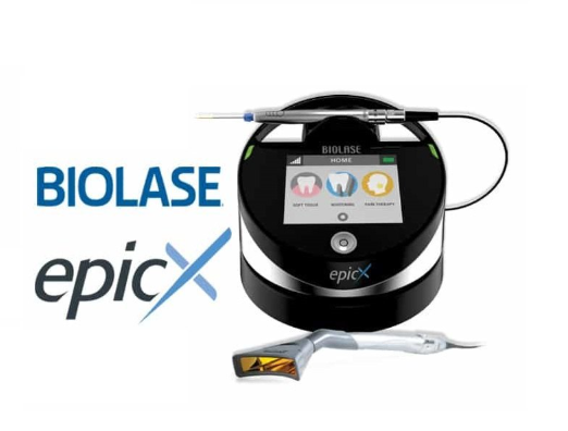
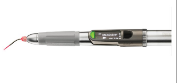
| BIOLASE Epic X | A soft-tissue laser shown in the original product illustration and listed by the manufacturer. [7] | The device picture does not identify the laser used in the cited trial or establish superiority for scanning. |
Practical review points before accepting a scan
The following are general review questions, not Amosso’s protocol or a substitute for a patient-specific treatment plan.
- Record the intended margin position, tissue condition and reason displacement is needed.
- Identify the exact technique, product or device and consult its instructions, contraindications and safety requirements.
- Check whether the field and finish line can be assessed after the intervention, rather than assuming displacement has been successful.
- Review the captured margin and relevant scan surfaces. If the record remains inadequate, reassess the clinical situation instead of accepting a complete-looking model as proof.
- Assess tissue response and the delivered restoration separately from the scanning result.
Original clinical and product illustrations
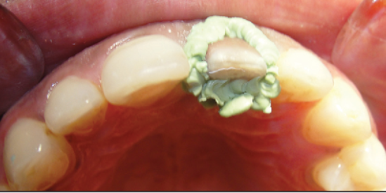
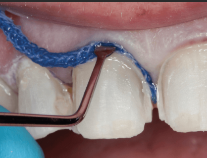
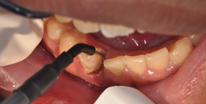
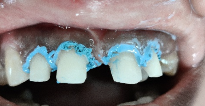
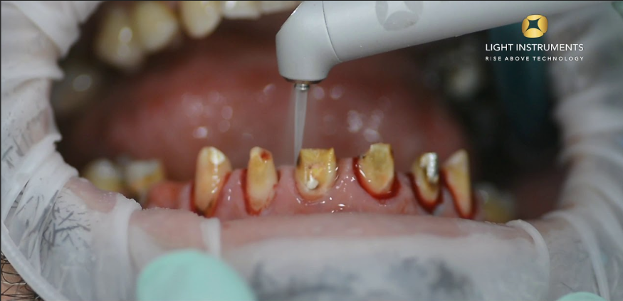
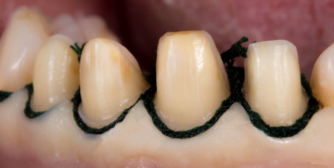
These retained images illustrate the subject of the article. They are not presented as a documented case series, before-and-after results, or verified steps of Amosso’s technique. Their original clinical histories and image permissions were not established by the sources reviewed here; no new clinical outcome is inferred from them.
Clinical application and remaining uncertainty
For a single crown, the useful question is whether the chosen approach provides a recordable margin with an acceptable tissue response in that patient. For a multi-unit or full-arch record, the premolar trial cannot settle all acquisition and fit questions. These are decision contexts, not reported patient cases. The earlier outline did not supply case records, outcome data or attributable testimonials, so none are claimed here.
Future evaluations should report the population, tissue condition, intervention, scanner and follow-up, and distinguish displacement, comfort, tissue change and restorative outcomes. Claims about AI-guided displacement or new biomaterials require identifiable evidence before being treated as established clinical capabilities.
Conclusion
Select gingival-displacement methods through clinical assessment and product-specific guidance, then verify the tissue response and scan separately. The available comparative evidence shows an access-versus-tissue tradeoff rather than a universal best method. Amosso’s original technique remains attributed and linked, with its unreviewed procedural details left to the source.
References and manufacturer resources
- Amosso E. A technique for gingival displacement before digital scanning. J Prosthet Dent. 2025;134(6):2636–2637. Online November 8, 2024. DOI; PubMed. Bibliographic identity verified; full technique not reviewed.
- El Ashry MF, et al. The efficacy of different gingival displacement methods for definitive digital impressions: A randomized controlled trial. J Dent. 2025;159:105841. PubMed abstract; DOI.
- Influence of the Gingival Condition on the Performance of Different Gingival Displacement Methods—A Randomized Clinical Study. J Clin Med. 2021;10(13):2747. Open-access primary study. DOI: 10.3390/jcm10132747.
- ACTEON. Manufacturer resource for Expasyl and Expasyl Exact. Product identification, not independent comparative evidence.
- Premier Dental. Traxodent retraction paste resource and manufacturer website. Promotional claims are not adopted as comparative findings.
- Premier Dental. Traxodent hemostatic gel introduction, August 8, 2025. Distinct gel variants; consult the applicable instructions.
- BIOLASE. Manufacturer website and soft-tissue laser resources. Device identification and instructions, not clinical superiority evidence.